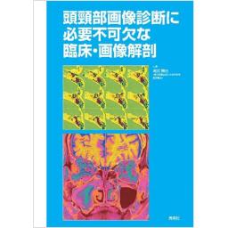
頭頸部画像診断に必要不可欠な臨床・画像解剖

出版社: Gakken(旧:学研メディカル秀潤社)
著者:
発行日: 2015-04-25
分野: 臨床医学:一般  >  画像/超音波
ISBN: 9784780909111
電子書籍版: 2015-04-01 (電子版Version1.0)
書籍・雑誌
≪全国送料無料でお届け≫
取寄せ目安:8~14営業日

5,610 円(税込)

電子書籍
章別単位での購入はできません
ブラウザ、アプリ閲覧

5,610 円(税込)

特記事項:
  • 【電子書籍】著作権者の意向により電子版では非掲載の画像がございます

商品紹介

好評だった画像診断2011年臨時増刊号を新装書籍化!一般のアトラス的要素の強い解剖書に対し,本書では頭頸部において臨床上重要と判断される代表的な解剖学的構造をスポット的に取り上げ,臨床解剖,画像解剖とともに臨床的意義を強調しました.

目次

  • 頭頸部画像診断に必要不可欠な臨床・画像解剖

    ―目次―

    1.眼窩
    2.鼻腔・副鼻腔の正常解剖
    3.鼻腔・副鼻腔の正常変異
    4.脳神経
    5.頭蓋底
    6.側頭骨(鼓室および顔面神経)
    7.側頭骨(内耳、内耳道)
    8.耳下腺
    9.上咽頭
    10.中咽頭
    11.口腔、口腔底
    12.下咽頭・喉頭領域
    13.頸部軟部組織・深部組織間隙
    14.頸部リンパ節

この書籍の参考文献

参考文献のリンクは、リンク先の都合等により正しく表示されない場合がありますので、あらかじめご了承下さい。

本参考文献は電子書籍掲載内容を元にしております。

1 眼窩

P.20 掲載の参考文献
1) Mafee MF : Orbit and visual pathways. The eye, Orbit : embryology, anatomy and pathology. In Som PM, Curtin HD (eds) ; Head and neck imaging. 4th ed, Mosby, St. Louis, p.439-654, 2003.
2) 酒井修 : 眼窩 眼窩・眼球の解剖. 多田信平, 黒崎喜久 (編) ; 頭頸部のCT・MRI. メディカル・サイエンス・インターナショナル, p.122-178, 2002.
3) 藤田晃史, 酒井修 : 眼窩. 酒井修 (編) ; 頭頸部の画像診断. 秀潤社, p.134-169, 2002.
4) Moore KL, Dalley AF : Orbit. In Moore KL, Dalley AF (eds) ; Clinically oriented anatomy. 4th ed, Lippincott Williams & Wilkins, Baltimore, p.899-915, 1999.
18) Lirng JF, Fuh JL, Wu ZA, et al : Diameter of the superior ophthalmic vein in relation to intracranial pressure. AJNR 24 : 700-703, 2003.
19) Mafee MF, Peyman GA : Retinal and choroidal detachments : role of magnetic resonance imaging and computed tomography. Radiol Clin North Am 25 : 487-507, 1987.
21) Peyman GA, Mafee MF : Uveal melanoma and similar lesions : the role of magnetic resonance imaging and computed tomography. Radiol Clin North Am 25 : 471-486, 1987.
22) 酒井修, 田村和哉, 田中修・他 : ぶどう膜悪性黒色腫のMRI-US, CTおよび病理所見との比較を中心に. 臨床放射線 37 : 207-212, 1992.
2 鼻腔・副鼻腔の正常解剖

P.38 掲載の参考文献
3) Pernkopf E (著) : Ferner H (編) ; 小川鼎三, 石川浩一 (訳) ; 臨床応用局所解剖図譜. 医学書院, 1966.
4) 上條雍彦 : 1 骨学. 口腔解剖学. アナトーム社, p.76-98, 1965.
6) Zinreich SJ, Benson ML, Oliverio PJ : Sinonasal cavities : CT normal anatomy, imaging of the osteometal complex, and functional endoscopic surgery. In Som PM, Curtin HD (ed) ; Head and neck imaging. 2nd ed, Mosby, St Louis, p.97-125, 1996.
7) Som PM, Brandwein M : Sinonasal cavities : anatomy, physiology, and plain film normal anatomy. In Som PM, Curtin HD (ed) ; Head and neck imaging. 4th ed, Mosby, St Louis, 2003.
8) Wallace R, Salazar JE, Cowles S : The relationship between frontal sinus drainage and osteomeatal complex disease : a CT study in 217 patients. AJNR 11 : 183-186, 1990.
9) Daniels DL, Mafee MF, Smith MM, et al : The frontal sinus drainage pathway and related structures. AJNR 24 : 1618-1627, 2003.
12) 池田勝久 : 内視鏡的副鼻腔手術のための臨床解剖. 池田勝久, 加我君孝, 岸本誠司・他 (編) ; 耳鼻咽喉科診療プラクティス8 耳鼻咽喉科・頭頸部外科のための臨床解剖. 文光堂, p.126-131, 2002.
13) 豊田圭子 : 鼻副鼻腔の解剖. 多田信平, 黒崎喜久 (編) ; 頭頸部のCT・MRI. メディカル・サイエンス・インターナショナル, p.180-189, 2002.
14) 大西俊郎 : ESSのための鼻腔, 副鼻腔の解剖. 大西俊郎, 小澤仁, 笠原行喜・他 (編) ; 内視鏡的副鼻腔手術. メジカルビュー社, p.32-45, 1996.
15) Naumann HH : Pathologishe anatomie der chronischen rhinitis und sinutitis. Proceedings VIII International Congress of Oto-Rhino-Laryngology. p.79, 1965.
17) Yonetsu K, Watanabe M, Nakamura T : Age-related expansion and reduction in aeration of the sphenoid sinus : volume assessment by helical CT scanning. AJNR 21 : 179-182, 2000.
19) 尾尻博也, 井田正博, 多田信平 : 頭蓋・顔面の骨折. 画像診断 17 : 599-607, 1997.

3 鼻腔・副鼻腔の正常変異

P.53 掲載の参考文献
1) 大西俊郎 : Chapter 3 FESSの理論. 大西俊郎, 小澤仁, 笠原行喜・他 (編) ; 内視鏡的副鼻腔手術. メジカルビュー社, p.17-18, 1995.
7) Kubal WS : Sinonasal anatomy. Neuroimaging Clin N Am 8 : 143-156, 1998.
9) Zinreich SJ, Benson ML, Oliverio PJ : Sinonasal cavities : CT normal anatomy, imaging of the osteomeatal comples, and functional endoscopic surgery. In Som PM, Curtin HD (eds) ; Head and neck imaging. 3rd ed, Mosby, St. Louis, p.97-125, 1996.
10) Lim CC, Dillon WP, McDermott MW : Mucocele involving the anterior clinoid process : MR and CT findings. AJNR 20 : 287-290, 1999.
13) 尾尻博也 : 2章 鼻副鼻腔. 頭頸部の臨床画像診断学. 第2版, 南江堂, p.43-54, 2011.
14) Mafee MR, Chow JM, Meyers R : Imaging anatomy of nasal cavity and paranasal sinuses for endoscopic sinus sugery. In Neuroradiology ARRS Categorical Courses Syllabus. American Collate of Radiology, Reston, 1992.

4 脳神経

P.69 掲載の参考文献
4) Yousry I, Moriggl B, Schmid UD, et al : Trigeminal ganglion and its divisions : detailed anatomic MR imaging with contrast-enhanced 3D constructive interference in the steady state sequences. AJNR 26 : 1128-1135, 2005.
5) Yagi A, Sato N, Taketomi A, et al : Normal cranial nerves in the cavernous sinuses : contrast-enhanced three-dimensional constructive interference in the steady state MR imaging. AJNR 26 : 946-950, 2005.

5 頭蓋底

P.86 掲載の参考文献
2) Janfaza P, Joseph BN : Scalp, cranium, and brain. In Surgical anatomy of the head and neck. Lippincott Williams & Wilkins, Philadelphia, p.49-147, 2001.
5) Li C, Yousem DM, Hayden RE, et al : Olfactory neuroblastoma : MR evaluation. AJNR 14 : 1167-1171, 1993.
7) 宜保浩彦, 外間政信, 大沢道彦 : 海綿静脈洞. 小林茂昭 (編) ; 臨床のための脳局所解剖学. 中外医学社, p.88-101, 2000.
15) Curtin HD : Detection of perineural spread : fat is a friend. AJNR 19 : 1385-1386, 1998.
19) Fruin ME, Smoker WR, Harnsberger HR : The carotid space in the suprahyoid neck. Semin Ultrasound CT MR 11 : 504-519, 1990.

6 側頭骨 ( 鼓室および顔面神経 )

P.107 掲載の参考文献
3) Mafee MF : MRI and CT in the evaluation of acquired and congenital cholesteatomas of the temporal bone. J Otolaryngol 22 : 239-248, 1993.
4) Weissman JL, Hirsch BE : Beyond the promontory : the multifocal origin of glomus tympanicum tumors. AJNR 19 : 119-122, 1998.
6) Thiers FA, Sakai O, Poe DS, et al : Persistent Stapedial Artery : CT Findings. AJNR 21 : 1551-1554, 2000.
7) 藤井直子, 片田和廣, 吉岡哲志・他 : マルチスライスCTによるCT基準面に対する耳小骨の角度計測と高分解能多断面再構成を用いた耳小骨最適断面の作製法. Otol Jpn 15 : 625-632, 2005.
9) 松本滋, 戸崎光宏, 宮崎日出海 : 耳小骨と耳小骨靱帯のMultislice CT : 正常解剖の描出と耳小骨奇形の診断. 画像医学誌 20 : 133-144, 2001.
12) Chadwell JB, Halsted MJ, Choo DI, et al : The cochlear cleft. AJNR 25 : 21-24, 2004.
18) Sartoretti-Schefer S, Brandle P, Wichmann W, et al : Intensity of MR contrast enhancement does not correspond to clinical and electro neurographic findings in acute infalammatory facial nerve palsy. AJNR 17 : 1229-1236, 1996.
24) Muckle RP, De la Cruz A, Lo WM : Petrous apex lesions. Am J Otol 19 : 219-225, 1998.

7 側頭骨 ( 内耳, 内耳道 )

P.118 掲載の参考文献
5) Chadwell JB, Halsted MJ, Choo DI, et al : The cochlear cleft. AJNR 25 : 21-24, 2004.
10) 長縄慎二 : 内耳の造影MRIによる内リンパ水腫画像-その撮像と評価について, いま放射線科医が知っておくべきこと- . 画像診断 35 : 278-289, 2015.

8 耳下腺

P.129 掲載の参考文献
1) Cross RR, Shapiro MD, Som PM : Parotid gland : MRI of the parapharyngeal space. Radiol Clin North Am 27 : 364-365, 1989.
2) Som PM, Brandwein MS : Parotid gland, salivary glands : anatomy and pathology. In Som PM, Curtin HD (eds) ; Head and Neck Imaging. 4th ed, Mosby, St. Louis, p.2007-2010, 2003.
5) Dailiana T, Chakeres D, Schmalbrock P, et al : High-resolution MR of the intraparotid facial nerve and parotid duct. AJNR 18 : 165-172, 1997.
9) Shimizu K, Iwai H, Ikeda K, et al : Intraparotid facial nerve schwannoma : a report of five cases and an analysis of magnetic resonance imaging results. AJNR 26 : 1328-1330, 2005.
12) Ikeda K, Kuroda M, Komemushi A, et al : The location of parotid gland tumors : evaluation with the main parotid duct on T2-weighted MRI and MR-sialography compared with other anatomical landmarks. 89th annual meeting of RSNA 2003, Chicago.
13) Moon WK, Han MH, Kim IO, et al : Congenital fistula from ectopic accessory parotid gland : diagnosis with CT sialography and CT fistulography. AJNR 16 : 997-999, 1995.
14) Hamilton BE, Saltzman KL, Wiggins RH 3rd, et al : Earring lesions of the parotid tail. AJNR 24 : 1757-1764, 2003.

9 上咽頭

P.142 掲載の参考文献
1) 西村龍一 : 5. 上咽頭. 高橋睦正 (編著) ; 頭頸部画像診断学. 中外医学社, p.94, 1997.
3) Hansberger, HR : Handbook of head and neck imaging. 2nd ed, Mosby, St. Louis, 1995.
4) Lentz M : Paranasal sinusitis and nasopharynx. In CT and MRI of head and neck tumors : methods, guidelines, differential diagnosis and clinical results. Thieme, New York, p.28-64, 1993.
7) Mancuso AA, Hanafee WN : Nasopharynx and parapharyngeal space. In Computed tomography and magnetic resonance imaging of the head and neck. 2nd ed, Lippincott Williams & Wilkins, Baltimore, p.428-498, 1985.
9) Curtin HD : Detection of perinueral spread : fat is a friend. AJNR 19 : 1385-1386, 1998.
13) 尾尻博也 : 8章 頸部リンパ節転移. 頭頸部の臨床画像診断学. 第2版, 南江堂, p.320, 2011.
16) 尾尻博也 : 3章 上咽頭. 頭頸部の臨床画像診断学. 第2版, 南江堂, p.117-137, 2011.
17) 岡村光英, 小山孝一 : 第4章 腫瘍. クリニカルPETの最前線. 先端医療技術研究所, p.76-82, 2004.

10 中咽頭

P.157 掲載の参考文献
1) 岸本誠司 : 頭頸部扁平上皮癌画像診断に必要な臨床的知識. 画像診断 22 : 1124-1132, 2002.
3) Kimura Y, Sumi M, Sumi T, et al : Deep extension from carcinoma arising from the gingiva : CT and MR imaging features. AJNR 23 : 468-472, 2002.
4) Mukherji SK, Holliday RA : Pharynx. In Som PM, Curtin HD (eds) ; Head and neck imaging. 3rd ed, Mosby, St. Louis, p.437-472, 1996.
5) Yonetsu K, Izumi M, Nakamura T : Deep facial infections of odontogenic origin : CT assessment of pathways of space involvement. AJNR 19 : 123-128, 1998.

11 口腔, 口腔底

P.175 掲載の参考文献
1) 上條雍彦 : 口腔解剖学. 第5巻 内臓学. 第1版, アナトーム社, p.1277, 1986.
2) Smoker WRK : Chapter 27. The oral cavity. In Som PM, Curtin HD (eds) ; Head and neck imaging Vol 2. 4th ed, Mosby, St. Louis, p.1377-1464, 2003.
3) 辰野聡 : 中咽頭・口腔の正常解剖. 多田信平, 黒崎喜久 (編) ; 頭頸部のCT・MRI. 第1版, メデイカル・サイエンス・インターナショナル, p.410-446, 2002.
4) Macdonald AJ, Harnsberger HR : Section 4. Oral Cavity. In Harnsberger HR (ed) ; Diagnostic imaging head and neck. AMIRSYS, Utah, III-4. 2-3, 2006.
5) Gale RD : Chapter 20. CT and MRI of the oral cavity and oropharynx. In Valvassori GE, Mafee MF, Carter BL (eds) ; Imaging of the head and neck. Thieme, New York, p.445-474, 1995.
8) 木村幸紀, 柳澤昭夫, 鎌田信悦・他 : 舌癌切除後のリンパ節転移の予知因子について-舌部分切除単独治療症例を用いての検討. 頭頸部腫瘍 22 : 78-82, 1996.
10) 日本頭頸部癌学会 (編) ; 頭頸部癌取扱い規約. 第5版, 金原出版, p.32-34, 2012.
11) Murakami R, Baba Y, Nishimura R, et al : CT and MR findings of denervated tongue after radical neck dissection. AJNR 18 : 747-750, 1997.
12) 上條雍彦 : 口腔解剖学. 第3巻 脈管学. 第1版, アナトーム社, p.446, 552, 555, 付22, 1986.
13) 上條雍彦 : 口腔解剖学. 第4巻 神経学. 第2版, アナトーム社, p.889-890, 895, 898-899, 963, 1987.
14) 木村幸紀, 花澤智美, 岡野友宏 : 画像所見から舌癌および口底癌の舌リンパ節転移が示唆された症例の臨床的検討と文献的考察. 頭頸部癌 36 : 488-497, 2010.
15) 上條雍彦 : 口腔解剖学. 第2巻 筋学. 第2版, アナトーム社, p.233, 290, 357, 1987.
16) 木村幸紀, 佐野, 岡野友宏 : 歯性感染症例に生じた頸動脈鞘膿瘍-CT像からみた波及経路の検討. 歯科放射線 40 : 38-46, 2000.
18) 木村幸紀, 花澤智美 , 岡野友宏 : 頬リンパ節・下顎リンパ節転移症例の検討. 歯科放射線 39 : 208-217, 2000.
21) 上條雍彦 : 口腔解剖学. 第1巻 骨学. 第2版, アナトーム社, p.75, 96, 150-155, 1987.
23) Kimura Y, Hanazawa T, Okano T, et al : Lateral retropharyngeal node metastasis from carcinoma of the upper gingiva and maxillary sinus. AJNR 19 : 1221-1224, 1998.
24) Ginberg LE, DeMonte F : Imaging of perineural tumor spread from palatal carcinoma. AJNR 19 : 1417-1422, 1998.

12 下咽頭・喉頭領域

P.194 掲載の参考文献
1) Mancuso AA : Evaluation and staging of laryngeal and hypopharyngeal cancer by computed tomography and magnetic resonance imaging. In Silver CE (ed) ; Laryngeal cancer. Thieme, New York, 1991.
9) 日本頭頸部腫瘍学会 (編) ; 頭頸部癌取扱い規約. 第5版, 金原出版, 2012.
13) Hiebert CA : Recurrent laryngeal nerve palsy. In Pearson FG, Deslauriers J, Ginsberg RJ, et al (eds) ; Thoracic surgery. Churchill Libingstone, New York, p.277-284, 1995.
14) Orringer MB, Orringer JS : Esophagectomy without thoracotomy : a dangerous operation? J Thorac Cardiovasc Surg 85 : 72-80, 1983.
15) Orringer MB : Complications of esophageal surgery. In Orringer MB, Zuidema GD (eds) ; Shackerford's surgery of the alimentary tract. Vol.1, The esophagus. 3rd ed, Saunders, Philadelphia, p.434-459, 1991.

13 頸部軟部組織・深部組織間隙

P.209 掲載の参考文献
1) 佐藤達夫 : 頭頸部外科に必要な局所解剖 (3) 頸部の筋膜. 耳喉頭頸 65 : 181-188, 1993.
4) Lemmon ML : Superficial fascia rhytidectomy. A restoration of the SMAS with control of the cervicomental angle. Clin Plast Surg 10 : 449-478, 1983.
9) 長谷川泰久 : 頸部郭清術のための臨床解剖. 池田勝久, 加我君孝, 岸本誠司・他 (編) ; 耳鼻咽喉科診療プラクティス8 耳鼻咽喉科・頭頸部外科のための臨床解剖. 文光堂, p.198-204, 2002.
10) 尾尻博也 : 第8章 頸部リンパ節転移. 頭頸部の臨床画像診断学. 第2版, 南江堂, p.309-351, 2011.
11) Rouviere H : Lymphatic system of the head and neck. In Tobias MJ (trans) ; Anatomy of the human lymphatic system. Edwards Brothers, Ann Arbor, p.5-28, 1938.
16) Eastwood JD, Hudgins PA, Malone D : Retropharyngeal effusion in acute calcific prevertebral tendonitis ; diagnosis with CT and MR imaging. AJNR 19 : 1789-1792, 1998.
17) Harnsberger HR : The perivertebral space. In Harnsberger HR (ed) ; Handbook of head and neck imaging. 2nd ed, Mosby, St Louis, p.105-119, 1995.

14 頸部リンパ節

P.227 掲載の参考文献
1) Mancuso AA : Infrahyoid neck, head and neck lymphatic system, and thoracic inlet : introduction and general principles. In Mancuso AA, Hanafee WN (eds) ; Head and neck radi o logy. Lippincott Williams & Wilkins, Philadelphia, p.1189-1217, 2010.
3) Som PM, Brandwein MS : Lymph nodes. In Som PM, Curtin HD (eds) ; Head and neck imaging. 4th ed, Mosby, St. Louis, p.1865-1934, 2003.
6) Rouviere H : Lymphatic system of the head and neck. In Tobias MJ (trans) ; Anatomy of the human lymphatic system. Edwards Brothers, Ann Arbor, p.5-28, 1938.
7) 尾尻博也 : 8章 頸部リンパ節転移. 頭頸部の臨床画像診断学. 第2版, 南江堂, p.309-351, 2011.
10) Mancuso AA, Mendenhall WM, Werning JW : Cervical metastatic disease and the "unknown" primary. In Mancuso AA, Hanafee WN (eds) ; Head and neck radiology. Lippincott Williams & Wilkins, Philadelphia, p.1294-1335, 2010.
12) 上條雍彦 ; 口腔解剖学. 3 脈管学 (基礎編). アナトーム社, 付1-36, 1991.

最近チェックした商品履歴

Loading...