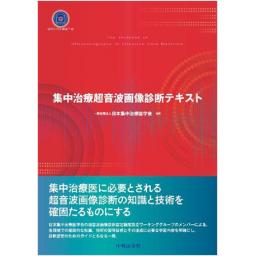
集中治療超音波画像診断テキスト

出版社: 中外医学社
著者:
発行日: 2023-03-10
分野: 臨床医学:一般  >  集中治療
ISBN: 9784498166509
電子書籍版: 2023-03-10 (1版1刷)
書籍・雑誌
≪全国送料無料でお届け≫
取寄せ目安:4~8営業日

5,500 円(税込)

電子書籍
章別単位で購入
ブラウザ、アプリ閲覧

5,500 円(税込)

商品紹介

ベッドサイドで医師自らが施行する point-of-care ultrasonography(POCUS)は重症患者をICU外へ移動させる必要がなく,集中治療領域でもその有用性が高まっている.本書は集中治療の現場で必要な知識や技術の習得のため,総論では超音波の基礎,各論では心・肺・血管・腹部・脳神経など全身をカバーし,小児エコーとともにショック状態のエコー診断を主要テーマとし解説した教科書である.

目次

  • I 総 論
     1 超音波診断(検査)の基礎
      1 超音波診断の基礎を学ぶ意義
      2 音響工学の知識
      3 プローブとオリエンテーション
      4 超音波装置の取り扱い
      5 アーチファクト
      6 超音波診断と医療安全

    II 各 論
     1 経胸壁心エコー
      1 経胸壁心エコー総論
      2 描出方法
      3 ICU 心エコーにおける各エコー所見から想定できる重要な病態
      4 ICU 心エコーで推定できる重要な臨床病態とエコー所見
     2 肺エコー
      1 肺エコー総論
      2 肺エコー各論
     3 横隔膜エコー
      1 横隔膜エコー総論
      2 横隔膜エコー各論
     4 気道エコー
      1 気道エコー総論
      2 気道エコー各論
     5 胃エコー
      1 胃エコー総論
     6 頸動脈エコー
      1 頸動脈エコー総論
      2 頸動脈エコー各論
     7 腹部・消化器エコー
      1 腹部・消化器エコー総論
      2 腹腔内出血,腹水貯留
      3 腸管麻痺
      4 胆嚢炎・胆管拡張
      5 水腎症
      6 腹部大動脈瘤・大動脈解離
     8 脳神経エコー TC-CFI
      1 脳神経エコー総論
      2 TC-CFI のエコーウインドウと描出方法
      3 脳神経エコーの実臨床への応用,得られた画像と計測値の解釈(MCA-MFV とLR)
      4 脳神経エコーの実臨床への応用,得られた画像と計測値の解釈(PI とONSD)
     9 下肢血管エコー
      1 下肢深部静脈血栓の評価
     10 ショック
      1 閉塞性ショック
      2 循環血漿量減少性ショック
      3 心原性ショック
      4 血液分布異常性ショック
      5 輸液反応性【アドバンス領域】
     11 小児エコー
      1 小児エコー総論
      2 心エコー
      3 声帯エコー
      4 肺エコー
      5 横隔膜エコー
      6 ショック
      7 神経超音波

この書籍の参考文献

参考文献のリンクは、リンク先の都合等により正しく表示されない場合がありますので、あらかじめご了承下さい。

本参考文献は電子書籍掲載内容を元にしております。

I 総論

P.21 掲載の参考文献
1) 亀田徹, 第1章 Point-of-Care 超音波の基本, 内科救急で使える! Point of care 超音波ベーシックス. 東京 : 医学書院 ; 2019. p.8-31.
2) Merit CRB. Physics of Ultrasound. Rumack CM, et al, editors. Diagnostic ultrasound 4th ed. Philadelphia : Elsevier ; 2011. p.1-33.
3) Wells PN. Ultrasonics in clinical diagnosis. Review Sci Basis Med Annu Rev. 1966 ; 38-53.
4) 神山直久, 亀田徹, 紺野啓, ほか. 第2章 超音波検査の基礎. 亀田徹, 木村昭夫, 編. 救急超音波テキスト. 東京 : 中外医学社 ; 2018. p.28-63.
5) AIUM technical bulletin. Transducer manipulation. American institute of ultrasound in medicine. J Ultrasound Med. 1999 ; 18 : 169-75.
6) Hedrick WR, Hykes DL, Starchman DE. Image artifacts. Hedrick WR, Hykes DL, Starchman DE, et al, editors. Ultrasound physics and instrumentation. St. Louis : Elsevier Mosby ; 2005. p.183-96.
8) Kameda T, Kamiyama N, Kobayashi H, et al. Ultrasonic B-line-like artifacts generated with simple experimental models provide clues to solve key issues in B-Lines. Ultrasound Med Biol. 2019 ; 45 : 1617-26.
10) 日本超音波医学会. 超音波診断装置の安全性に関する資料 第4版. https://www.jsum.or.jp/committee/uesc/pdf/safty.pdf (2021年2月28日アクセス)
11) U.S. FDA. Marketing clearance of diagnostic ultrasound systems and transducers, guidance for industry and Food and Drug Administration staff (2019/6/27). https://www.fda.gov/media/71100/download (2021年2月28日アクセス)
12) Guideline for ultrasound transducer cleaning and disinfection. Ann Emerg Med. 2018 ; 72 (4) : e45-e47.
13) American Institute of Ultrasound in Medicine. Quick Guide on COVID-19 Protections-Ultrasound Transducers, Equipment, and Gel. https://www.jsum.or.jp/committee/uesc/pdf/covid19_quick_guide.pdf (2021年2月28日アクセス)
14) 医用超音波用語集. 日本超音波医学会ホームページ. https://www.jsum.or.jp/terminologies (2021年4月4日アクセス)
II 各論

P.35 掲載の参考文献
2) Via G, Hussain A, Wells M, et al. International evidence-based recommendations for focused cardiac ultrasound. J Am Soc Echocardiogr. 2014 ; 27 : 683.
2) Sievers B, Kirchberg S, Franken U, et al. Visual estimation versus quantitative assessment of left ventricular ejection fraction : a comparison by cardiovascular magnetic resonance imaging. Am Heart J. 2005 ; 150 : 737-42.
3) Fowler NO. Cardiac tamponade. A clinical or an echocardiographic diagnosis? Circulation. 1993 ; 87 : 1738-41.
3) Levitov A, Frankel HL, Blaivas M, et al. Guidelines for the appropriate use of bedside general and cardiac ultrasonography in the evaluation of critically ill patients-part II : cardiac ultrasonography. Crit Care Med. 2016 ; 44 : 1206-27.
4) Neskovic AN, Skinner H, Price S, et al. Focus cardiac ultrasound core curriculum and core syllabus of the European Association of Cardiovascular Imaging. Eur Heart J Cardiovasc Imaging. 2018 ; 19 : 475-81.
5) Vieillard-Baron A, Millington SJ, Sanfilippo F, et al. A decade of progress in critical care echocardiography : a narrative review. Intensive Care Med. 2019 ; 45 : 770-88.
6) Wong A, Galarza L, Forni L, et al. Recommendations for core critical care ultrasound competencies as a part of specialist training in multidisciplinary intensive care : a framework proposed by the European Society of Intensive Care Medicine (ESICM). Crit Care. 2020 ; 24 : 393.
P.45 掲載の参考文献
5) Lichtenstein D, Meziere G, Biderman P, et al. The comet-tail artifact. An ultrasound sign of alveolar-interstitial syndrome. Am J Respir Crit Care Med. 1997 ; 156 : 1640-6.
6) Lichtenstein D, Meziere G, Biderman P, et al. The comet-tail artifact : an ultrasound sign ruling out pneumothorax. Intensive Care Med. 1999 ; 25 : 383-8.
7) Yue Lee FC, Jenssen C, Dietrich CF. A common misunderstanding in lung ultrasound : the comet tail artefact. Med Ultrason. 2018 ; 20 : 379-84.
8) Idei M, Seino Y, Nomura T. Pseudo B-line in pneumothorax. Intensive Care Med. 2020 ; 46 : 1468-9.
9) Lichtenstein DA, Lascols N, Prin S, et al. The "lung pulse" an early ultrasound sign of complete atelectasis. Intensive Care Med. 2003 ; 29 : 2187-92.
10) Lichtenstein DA, Meziere G, Biderman P, et al. The "lung point" : an ultrasound sign specific to pneumothorax. Intensive Care Med. 2000 ; 26 : 1434-40.
11) Alrajab S, Youssef AM, Akkus NI, et al. Pleural ultrasonography versus chest radiography for the diagnosis of pneumothorax : review of the literature and meta-analysis. Crit Care. 2013 ; 17 : R208.
12) Abbasi S, Farsi D, Hafezimoghadam P, et al. Accuracy of emergency physician-performed ultrasound in detecting traumatic pneumothorax after a 2-h training course. Eur J Emerg Med. 2013 ; 20 : 173-7.
13) Lichtenstein D, Rialp G. Lung ultrasound in a dyspneic patient with giant bullae. Intensive Care Med. 2019 ; 45 : 690-1.
14) Volpicelli G, Boero E, Sverzellati N, et al. Semi-quantification of pneumothorax volume by lung ultrasound. Intensive Care Med. 2014 ; 40 : 1460-7.
15) 三澤賢治, 西智史, 西田保則, ほか. 超音波検査における自然気胸の重症度判定の試み. 日本呼吸器外科学会誌. 2010 ; 24 : 8-11.
16) Galbois A, Ait-Oufella H, Baudel JL, et al. Pleural ultrasound compared with chest radiographic detection of pneumothorax resolution after drainage. Chest. 2010 ; 138 : 648-55.
17) Soult MC, Collins JN, Novosel TJ, et al. Thoracic ultrasound can predict safe removal of thoracostomy tubes. J Trauma Acute Care Surg. 2014 ; 77 : 256-61.
P.49 掲載の参考文献
1) Verdon F, Herzig L, Burnand B, et al. Chest pain in daily practice : occurrence, causes and management. Swiss Med Wkly. 2008 ; 138 (23-24) : 340-7.
2) 三浦弘之, 平良修, 平栗俊介, ほか. 鈍的胸部外傷症例の検討, 日胸外会誌. 1998 ; 46 : 556-60.
3) Yousefifard M, Baikpour M, Ghelichkhani P, et al. Comparison of ultrasonography and radiography in detection of thoracic bone fractures ; a systematic review and meta-analysis. Emerg (Tehran). 2016 ; 4 (2) : 55-64.
4) Al-Thani H, Jabbour G, El-Menyar A, et al. Traumatic sternal injury in patients with rib fracture : a single-center experience. Int J Crit Illn Inj Sci. 2019 ; 9 (2) : 75-81.
5) You JS, Chung YE, Kim D, et al. Role of sonography in the emergency room to diagnose sternal fractures. J Clin Ultrasound. 2010 ; 38 (3) : 135-7.
6) Chen CL, Cheng YL. Delayed massive hemothorax complicating simple rib fracture associated with diaphragmatic injury. Am J Emerg Med. 2014 ; 32 : 818.e3-4.
7) Hills MW, Delprado AM, Deane SA. Sternal fractures : associated injuries and management. J Trauma. 1993 ; 35 : 55-60.
8) Montoya J, Stawicki SP, Evans DC, et al. From FAST to E-FAST : an overview of the evolution of ultrasound-based traumatic injury assessment. Eur J Trauma Emerg Surg. 2016 ; 42 (2) : 119-26.
P.51 掲載の参考文献
1) Kalokairinou-Motogna M, Maratou K, Paianid I, et al. Application of color Doppler ultrasound in the study of small pleural effusion. Med Ultrason. 2010 ; 12 : 12-6.
2) Lichtenstein D, Goldstein I, Mourgeon E, et al. Comparative diagnostic performances of auscultation, chest radiography, and lung ultrasonography in acute respiratory distress syndrome. Anesthesiology. 2004 ; 100 : 9-15.
3) Grimberg A, Shigueoka DC, Atallah AN, et al. Diagnostic accuracy of sonography for pleural effusion : systematic review. Sao Paulo Med J. 2010 ; 128 (2) : 90-5.
4) Eibenberger KL, Dock WI, Ammann ME, et al. Quantification of pleural effusions : sonography versus radiography. Radiology. 1994 ; 191 : 681-4.
5) Balik M, Plasil P, Waldauf P, et al. Ultrasound estimation of volume of pleural fluid in mechanically ventilated patients. Intensive Care Med. 2006 ; 32 : 318.
6) Vignon P, Chastagner C, Berkane V, et al. Quantitative assessment of pleural effusion in critically ill patients by means of ultrasonography. Crit Care Med. 2005 ; 33 : 1757-63.
7) Mayo PH, Goltz HR, Tafreshi M. Safety of ultrasound-guided thoracentesis in patients receiving mechanical ventilation. Chest. 2004 ; 125 : 1059-62.
8) Gordon CE, Feller-Kopman D, Balk EM, et al. Pneumothorax following thoracentesis : a systematic review and meta-analysis. Arch Intern Med. 2010 ; 170 : 332-9.
9) Mayo PH, Goltz HR, Tafreshi M, et al. Safety of ultrasound-guided thoracentesis in patients receiving mechanical ventilation. Chest. 2004 ; 125 : 1059-62.
10) Havelock T, Teoh R, Laws D, et al ; BTS Pleural Disease Guideline Group. Pleural procedures and thoracic ultrasound : British Thoracic Society Pleural Disease Guideline 2010. Thorax. 2010 ; 65 Suppl 2 : ii61-76.
P.53 掲載の参考文献
1) Nazerian P, Volpicelli G, Vanni S, et al. Accuracy of lung ultrasound for the diagnosis of consolidations when compared to chest computed tomography. Am J Emerg Med. 2015 ; 33 : 620-5.
2) Bourcier JE, Paquet J, Seinger M, et al. Performance comparison of lung ultrasound and chest x-ray for the diagnosis of pneumonia in the ED. Am J Emerg Med. 2014 ; 32 : 115-8.
3) Chavez MA, Shams N, Ellington LE, et al. Lung ultrasound for the diagnosis of pneumonia in adults : a systematic review and meta-analysis. Respir Res. 2014 ; 15 : 50.
4) Ye X, Xiao H, Chen B, et al. Accuracy of lung ultrasonography versus chest radiography for the diagnosis of adult community-acquired pneumonia : review of the literature and meta-analysis. PLoS One. 2015 ; 10 (6) : e0130066.
7) Lichtenstein D, Meziere G, Seitz J. The dynamic air bronchogram. A lung ultrasound sign of alveolar consolidation ruling out atelectasis. Chest. 2009 ; 135 : 1421-5.
P.55 掲載の参考文献
3) Volpicelli G, Mussa A, Garofalo G, et al. Bedside lung ultrasound in the assessment of alveolar-interstitial syndrome. Am J Emerg Med. 2006 ; 24 : 689-96.
4) Gargani L. Lung ultrasound : a new tool for the cardiologist. Cardiovasc Ultrasound. 2011 ; 9 : 6.
5) Copetti R, Soldati G, Copetti P. Chest sonography : a useful tool to differentiate acute cardiogenic pulmonary edema from acute respiratory distress syndrome. Cardiovasc Ultrasound. 2008 ; 6 : 16.
6) Reissig A, Kroegel C. Transthoracic sonography of diffuse parenchymal lung disease : the role of comet tail artifacts. J Ultrasound Med. 2003 ; 22 : 173-80.
7) Bouhemad B, Mongodi S, Via G, et al. Ultrasound for "lung monitoring" of ventilated patients. Anesthesiology. 2015 ; 122 : 437-47.
8) 川上大裕, 瀬尾隆太郎. 急性呼吸不全の鑑別診断 COPD 増悪 VS 心不全 特集 ICUエコー. Intensivist. 2017 ; 9 : 68-74.
9) Jambrik Z, Monti S, Coppola V, et al. Usefulness of ultrasound lung comets as a nonradiologic sign of extravascular lung water. Am J Cardiol. 2004 ; 93 : 1265-70.
P.58 掲載の参考文献
1) Matamis D, Soilemezi E, Tsagourias M, et al. Sonographic evaluation of the diaphragm in critically ill patients. Technique and clinical applications. Intensive Care Med. 2013 ; 39 : 801-10.
3) Jiang JR, Tsai TH, Jerng JS, et al. Ultrasonographic evaluation of liver/spleen movements and extubation outcome. Chest. 2004 ; 126 : 179- 85.
4) Boussuges A, Gole Y, Blanc P. Diaphragmatic motion studied by M-mode ultrasonography : methods, reproducibility, and normal values. Chest. 2009 ; 135 : 391-400.
5) Haji K, Royse A, Green C, et al. Interpreting diaphragmatic movement with bedside imaging, review article. J Crit Care. 2016 ; 34 : 56-65.
6) Zambon M, Greco M, Bocchino S, et al. Assessment of diaphragmatic dysfunction in the critically ill patient with ultrasound : a systematic review. Intensive Care Med. 2017 ; 43 : 29-38.
7) Goligher EC, Heunks LMA, Brochard LJ. Critical illness-associated diaphragm weakness. Intensive Care Med. 2017 ; 43 : 1441-52.
8) Goligher EC, Dres M, Fan E, et al. Ferguson mechanical ventilation-induced diaphragm atrophy strongly impacts clinical outcomes. Am J Respir Crit Care Med. 2018 ; 197 : 204-13.
9) Goligher EC, Fan E, Herridge MS, et al. Ferguson evolution of diaphragm thickness during mechanical ventilation impact of inspiratory effort. Am J Respir Crit Care Med. 2015 ; 192 : 1080-8.
10) Palkar A, Narasimhan M, Greenberg H, et al. Diaphragm excursion-time index : a new parameter using ultrasonography to predict extubation outcome. Chest. 2018 ; 153 : 1213-20.
P.64 掲載の参考文献
2) Kameda T, Fujita M. Point-of-care ultrasound detection of tracheal wall thickening caused by smoke inhalation. Crit Ultrasound J. 2014 ; 6 : 11.
3) Suzuki A, Iida T, Kunisawa T, et al. Ultrasound-guided cannula cricothyroidotomy. Anesthesiology. 2012 ; 117 : 1128.
4) 鈴木昭広, 稲垣泰好, 後藤祐也, ほか. 超音波によるプレスキャンを利用した安全な経皮的気管切開術の試み. 日本集中治療学会雑誌. 2013 ; 20 : 293-4.
P.68 掲載の参考文献
1) El-Boghdadly K, Wojcikiewicz T, Perlas A. Perioperative point-of-care gastric ultrasound. BJA Educ. 2019 ; 19 (7) : 219-26.
P.73 掲載の参考文献
1) 厚生労働省. 令和元年 (2019) 人口動態統計月報年計 (概数) の概況. https://www.mhlw.go.jp/toukei/saikin/hw/jinkou/geppo/nengai19/dL/gaikyouR1.pdf (2021年2月閲覧)
2) NASCET collaborators. Beneficial effect of carotid endarterectomy in symptomatic patients with high-grade carotid stenosis. N Engl J Med. 1991 ; 325 : 445-53.
3) McColgan P, Bentley P, McCarron M, et al. Evaluation of the clinical utility of a carotid bruit. QJM. 2012 ; 105 : 1171-7.
5) Mita T, Katakami N, Yoshii H, et al. Alogliptin, a dipeptidyl peptidase 4 inhibitor, prevents the progression of carotid atherosclerosis in patients with type 2 diabetes : the study of preventive effects of alogliptin on diabetic Atherosclerosis (SPEAD-A). Diabetes Care. 2016 ; 39 : 139-48.
6) 早期動脈硬化研究会. http://www.imt-ca.com/ (2021年2月閲覧)
7) 日本超音波医学会用語・診断基準委員会. 頸動脈超音波診断ガイドライン小委員会 : 超音波による頸動脈病変の標準的評価法 2017.
8) Wu X, Studer W, Skarvan K, et al. High incidence of intravenous thrombi after short-term central venous catheterization of the internal jugular vein. J Clin Anesth. 1999 ; 11 : 482-5.
9) Baskin JL, Pui CH, Reiss U, et al. Management of occlusion and thrombosis associated with long-term indwelling central venous catheters. Lancet. 2009 ; 374 : 159-69.
P.101 掲載の参考文献
1) Blanco P, Blaivas M. Applications of transcranial color-coded sonography in the emergency department. J Ultrasound Med. 2017 ; 36 : 1251-66.
2) Lau VI, Arntfield RT. Point-of-care transcranial Doppler by intensivists. Crit Ultrasound J. 2017 ; 9 : 21.
3) T Gerriets, E Stolz, S Konig, et al. Sonographic monitoring of midline shift in space-occupying stroke : an early outcome predictor. Stroke. 2001 ; 32 : 442-7.
4) Liao C-C, Chen Y-F, Xiao F. Brain midline shift measurement and its automation : a review of techniques and algorithms. Int J Biomed Imaging. 2018 ; 2018 : 4303161.
5) Blanco P, Abdo-Cuza A. Transcranial Doppler ultrasound in neurocritical care. J Ultrasound. 2018 ; 21 : 1-16.
6) Kirsch JD, Mathur M, Johnson MH, et al. Advances in transcranial Doppler US : imaging ahead. Radiographics. 2013 ; 33 : E1-E14.
7) Francoeur CL, Mayer SA. Management of delayed cerebral ischemia after subarachnoid hemorrhage. Crit Care. 2016 ; 20 : 277.
8) Connolly ES Jr, Rabinstein AA, Carhuapoma JR, et al. Guidelines for the management of aneurysmal subarachnoid hemorrhage : a guideline for healthcare professionals from the American Heart Association/american Stroke Association. Stroke. 2012 ; 43 : 1711-37.
9) Diringer MN, Bleck TP, Hemphill JC 3rd, et al. Critical care management of patients following aneurysmal subarachnoid hemorrhage : recommendations from the Neurocritical Care Society's Multidisciplinary Consensus Conference. Neurocrit Care. 2011 ; 15 : 211-40.
10) Robba C, Taccone FS. How I use transcranial Doppler. Crit Care. 2019 ; 23 : 420.
11) Topcuoglu MA. Transcranial Doppler ultrasound in neurovascular diseases : diagnostic and therapeutic aspects. J Neurochem. 2012 ; 123 Suppl 2 ; 39-51.
12) Toi H, Matsumoto N, Yokosuka K, et al. Prediction of cerebral vasospasm using early stage transcranial Doppler. Neurol Med Chir (Tokyo). 2013 ; 53 : 396-402.
13) Munoz-Sanchez MA, Murillo-Cabezas F, Egea-Guerrero JJ, et al. Emergency transcranial doppler ultrasound : predictive value for the development of symptomatic vasospasm in spontaneous subarachnoid hemorrhage in patients in good neurological condition. Med Intensiva. 2012 ; 36 (9).
14) Sloan MA, Alexandrov AV, Tegeler CH, et al. Assessment : transcranial Doppler ultrasonography : report of the Therapeutics and Technology Assessment Subcommittee of the American Academy of Neurology. Neurology. 2004 ; 62 : 1468-81.
15) Tsivgoulis G, Alexandrov AV, Sloan MA. Advances in transcranial Doppler ultrasonography. Curr Neurol Neurosci Rep. 2009 ; 9 : 46-54.
16) Mastantuono J-M, Combescure C, Elia N, et al. Transcranial Doppler in the diagnosis of cerebral vasospasm : an updated meta-analysis. Crit Care Med. 2018 ; 46 : 1665-72.
17) Norisue Y, Fujimoto Y, Nakagawa K, et al. Preliminary guideline- and pathophysiology-based protocols for neurocritical care. J Intensive Care. 2018 ; 6 : 45.
18) Brain Trauma FOUNDATION. https://www.braintrauma.org
19) Goeres P, Zeiler FA, Unger B, et al. Ultrasound assessment of optic nerve sheath diameter in healthy volunteers. J Crit Care. 2016 ; 31 : 168-71.
20) Geeraerts T, Launey Y, Martin L, et al. Ultrasonography of the optic nerve sheath may be useful for detecting raised intracranial pressure after severe brain injury. Intensive Care Med. 2007 ; 33 : 1704-11.
21) Kimberly HH, Shah S, Marill K, et al. Correlation of optic nerve sheath diameter with direct measurement of intracranial pressure. Acad Emerg Med. 2008 ; 15 : 201-4.
22) Rajajee V, Vanaman M, Fletcher JJ, et al. Optic nerve ultrasound for the detection of raised intracranial pressure. Neurocrit Care. 2011 ; 15 : 506-15.
23) Soldatos T, Karakitsos D, Chatzimichail K, et al. Optic nerve sonography in the diagnostic evaluation of adult brain injury. Crit Care. 2008 ; 12 (3) : R67.
24) Agrawal D, Raghavendran K, Zhao L, et al. A prospective study of optic nerve ultrasound for the detection of elevated intracranial pressure in severe traumatic brain injury. Crit Care Med. 2020 ; 48 : e1278-e1285.
25) Bellner J, Romner B, Reinstrup P, et al. Transcranial Doppler sonography pulsatility index (PI) reflects intracranial pressure (ICP). Surg Neurol. 2004 ; 62 : 45-51 ; discussion 51.
26) Figaji AA, Zwane E, Fieggen AG, et al. Transcranial Doppler pulsatility index is not a reliable indicator of intracranial pressure in children with severe traumatic brain injury. Surg Neurol. 2009 ; 72 : 389-94.
27) Kristiansson H, Nissborg E, Bartek J Jr, et al. Measuring elevated intracranial pressure through noninvasive methods : a review of the literature. J Neurosurg Anesthesiol. 2013 ; 25 : 372-85.
P.108 掲載の参考文献
1) Tapson VF. Acute pulmonary embolism. N Engl J Med. 2008 ; 358 : 1037-52.
2) Ultrasound guidelines : emergency, point-of-care and clinical ultrasound guidelines in medicine. Ann Emerg Med. 2017 ; 69 (5) : e27-e54.
4) Pomero F, Dentali F, Borretta V, et al. Accuracy of emergency physician-performed ultrasonography in the diagnosis of deep-vein thrombosis : A systematic review and meta-analysis. Thromb Haemost. 2013 ; 109 : 137-45.
5) Needleman L, Cronan JJ, Lilly MP, et al. Ultrasound for lower extremity deep venous thrombosis. Circulation. 2018 ; 137 : 1505-15.
6) Shiloh AL, McPhee C, Eisen L, et al. Better with ultrasound : detection of DVT. Chest. 2020 ; 158 : 1122-7.
7) Lee JH, Lee SH, Yun SJ. Comparison of 2-point and 3-point point-of-care ultrasound techniques for deep vein thrombosis at the emergency department : a meta-analysis. Medicine (Baltimore). 2019 ; 98 : e15791.
8) Al-Khafaji RA, Schierbeck L. Deep venous thrombosis in a patient with a moderate pretest probability and a negative D-dimer test : a review of the diagnostic algorithms. J Blood Med. 2020 ; 27 (11) : 173-84.
9) Patel H, Sun H, Hussain AN, et al. Advances in the diagnosis of venous thromboembolism : a literature review. Diagnostics (Basel). 2020 ; 10 : 365.
10) Trischler T, Kraaijpoel N, Gal GL, et al. Venous thromboembolism : advances in diagnosis and treatment. JAMA. 2018 ; 320 : 1583-94.
11) Garcia JP, Alonso JV, Garcia PC, et al. Comparison of the accuracy of emergency department-performed point-of-care-ultrasound (POCUS) in the diagnosis of lower-extremity deep vein thrombosis. J Emerg Med. 2018 ; 54 : 656-64.
12) Patel P, Patel P, Bhatt M, et al. Systematic review and meta-analysis of outcomes in patients with suspected deep vein thrombosis. Blood advances. 2020 ; 4 : 2779-88.
14) Klok FA, Kruip MJHA, Meer NJM, et al. Incidence of thrombotic complications in critically ill ICU patients with COVID-19. Thromb Res. 2020 ; 191 : 145-7.
15) Cui S, Chen S, Li X, et al. Prevalence of venous thromboembolism in patients with severe novel coronavirus pneumonia. J Thromb Haemost. 2020 ; 18 : 1421-4.
16) Longchamp A, Longchamp J, Manzocci-Besson S, et al. Venous thromboembolism in critically Ill patients with COVID-19 : results of a screening study for deep vein thrombosis. Res Pract Thromb Haemost. 2020 ; 4 : 842-7.
17) Chen S, Zhang D, Zheng T, et al. DVT incidence and risk factors in critically ill patients with COVID-19. J Thromb Thrombolysis. 2020 ; 51 : 33-9.
P.119 掲載の参考文献
1) Rubano E, Mehta N, Caputo W, et al. Systematic review : emergency department bedside ultrasonography for diagnosing suspected abdominal aortic aneurysm. Acad Emerg Med. 2013 ; 20 : 128-38.
2) Unluer EE, Karagoz A, Akoglu H, et al. Visual estimation of bedside echocardiographic ejection fraction by emergency physicians. West J Emerg Med. 2014 ; 15 : 221-6.
3) Monnet X, Marik P, Teboul J-L. Passive leg raising for predicting fluid responsiveness : a systematic review and meta-analysis. Intensive Care Med. 2016 ; 42 : 1935-47.
4) Petersen SE, Aung N, Sanghvi MM, et al. Reference ranges for cardiac structure and function using cardiovascular magnetic resonance (CMR) in Caucasians from the UK Biobank population cohort. J Cardiovasc Magn Reson. 2017 ; 19 : 18.
5) Boyd JH, Forbes J, Nakada T, et al. Fluid resuscitation in septic shock : a positive fluid balance and elevated central venous pressure are associated with increased mortality. Crit Care Med. 2011 ; 39 : 259-65.
6) Wu Y, Zhou S, Zhou Z, et al. A 10-second fluid challenge guided by transthoracic echocardiography can predict fluid responsiveness. Crit Care. 2014 ; 18 (3) : R108.
7) Vignon P, Repesse X, Begot E, et al. Comparison of echocardiographic indices used to predict fluid responsiveness in ventilated patients. Am J Respir Crit Care Med. 2017 ; 195 : 1022-32.
9) Feissel M, Michard F, Faller J-P, et al. The respiratory variation in inferior vena cava diameter as a guide to fluid therapy. Intensive Care Med. 2004 ; 30 : 1834-7.
P.126 掲載の参考文献
1) Via G, Hussain A, Wells M, et al. International evidence-based recommendations for focused cardiac ultrasound. J Am Soc Echocardiogr. 2014 ; 27 : 683.e1-683.e33.
2) Levitov A, Frankel HL, Blaivas M, et al. Guidelines for the appropriate use of bedside general and cardiac ultrasonography in the evaluation of critically ill patients-part II : cardiac ultrasonography. Crit Care Med. 2016 ; 44 : 1206-27.
3) Rich S, Sheikh A, Gallastegui J, et al. Determination of left ventricular ejection fraction by visual estimation during real-time two-dimensional echocardiography. Am Heart J. 1982 ; 104 : 603-6.
4) Miller AF, Arichai P, Gravel CA, et al. Use of cardiac point-of-care ultrasound in the pediatric emergency department. Pediatr Emerg Care. 2022 ; 38 : e300-e305.
5) Alerhand S, Choi A, Valga P. Cardiac Ultrasound for Pediatric Emergencies. Pediatr Ann. 2021 ; 50 : e424-e431.
6) Levine AC, Shah SP, Umulisa I, et al. Ultrasound assessment of severe dehydration in children with diarrhea and vomiting. Acad Emerg Med. 2010 ; 17 : 1035-41.
P.127 掲載の参考文献
1) Garel C, Contencin P, Polonovski JM, et al. Laryngeal ultrasonography in infants and children : a new way of investigating. Normal and pathological findings. Int J Pediatr Otorhinolaryngol. 1992 ; 23 : 107-15.
2) Lee MGY, Millar J, Rose E, et al. Laryngeal ultrasound detects a high incidence of vocal cord paresis after aortic arch repair in neonates and young children. J Thorac Cardiovasc Surg. 2018 ; 155 : 2579-87.
3) Hamadah HK, Kabbani MS. Bedside ultrasound in the diagnosis and treatment of children with respiratory difficulty following cardiac surgery. J Clin Imaging Sci. 2017 ; 7 : 37.
4) Ongkasuwan J, Ocampo E, Tran B. Laryngeal ultrasound and vocal fold movement in the pediatric cardiovascular intensive care unit. Laryngoscope. 2017 ; 127 : 167-72.
5) 吉田美苗, 長谷川智巳, 金子尚樹, ほか. 小児大動脈弓再建 術後の声帯エコー評価. 日集中医誌. 2020 ; 27 : 104-8.
6) 金子尚樹, 長谷川智巳, 吉田美苗, ほか. 小児声帯麻痺に対する声帯エコー評価の有用性. 日集中医誌. 2020 ; 27 : 453-8.
P.128 掲載の参考文献
1) Lichtenstein DA, Mauriat P. Lung ultrasound in the critically ill neonate. Curr Pediatr Rev. 2012 ; 8 : 217-23.
2) Liu J, Copetti R, Sorantin E, et al. Protocol and guidelines for point-of-care & lung ultrasound in diagnosing neonatal pulmonary diseases based on international expert consensus. J Vis Exp 2019 ; (145). doi : 10.3791/58990. This is the first and only international expert consensus describing a protocol and guidelines for point-of-care lung ultrasound.
3) Orso D, Ban A, Guglielmo N. Lung ultrasound in diagnosing pneumonia in childhood : a systematic review and meta-analysis. J Ultrasound. 2018 ; 21 : 183-95.
P.130 掲載の参考文献
1) Sanchez de Toledo J, Munoz R, Landsittel D, et al. Diagnosis of abnormal diaphragm motion after cardiothoracic surgery : ultrasound performed by a cardiac intensivist vs. fluoroscopy. Congenit Heart Dis. 2010 ; 5 : 565-72.
2) Gil-Juanmiquel L, Grataco's M, Castilla-Ferna'ndez Y, et al. Bedside ultrasound for the diagnosis of abnormal diaphragmatic motion in children after heart surgery. Pediatr Crit Care Med. 2017 ; 18 : 159-64.
P.132 掲載の参考文献
1) Chen L, Hsiao A, Langhan M, et al. Use of bedside ultrasound to assess degree of dehydration in children with gastroenteritis. Acad Emerg Med. 2010 ; 17 : 1042-7.
2) Kosiak W, Swieton D, Piskunowicz M. Sonographic inferior vena cava/aorta diameter index, a new approach to the body fluid status assessment in children and young adults in emergency ultrasound-preliminary study. Am J Emerg Med. 2008 ; 26 : 320-5.
3) Park DB, Presley BC, Cook T, et al. Point-of-care ultrasound for pediatric shock. Pediatr Emerg Care. 2015 ; 31 : 591-8 ; quiz 599-601.
4) Weberding NT, Marin JR. Point-of-care ultrasound for targeted assessment of shock : remember HIPP in children. Pediatr Emerg Care. 2019 ; 35 : 575-8.
P.134 掲載の参考文献
1) Singh Y, Tissot C, Fraga MV, et al. International evidence-based guidelines on Point of Care Ultrasound (POCUS) for critically ill neonates and children issued by the POCUS Working Group of the European Society of Paediatric and Neonatal Intensive Care (ESPNIC). Cri Care. 2020 ; 24 : 65.
2) Lau VI, Arntfield RT. Point-of-care transcranial Doppler by intensivists. Crit Ultrasound J. 2017 ; 9 : 21.
3) Motuel J, Biette I, Srairi M, et al. Assessment of brain midline shift using sonography in neurosurgical ICU patinets. Crit Care. 2014 ; 18 : 676.
4) Ohle R, Mclsaac SM, Woo MY, et al. Sonography of the optic nerve sheath diameter for detection of raised intracranial pressure compared to computed tomography : a systematic review and meta-analysis. J Ultrasound Med. 2015 ; 34 : 1285-94.
5) Dubourg J, Javouhey E, Geeraerts T, et al. Ultrasonography of optic nerve sheath diameter for detection of raised intracranial pressure : a systematic review and meta-analysis. Intensive Care Med. 2011 ; 37 : 1059-68.
6) Malayeri AA, Bavarian S, Mehdizadeh M. Sonographic evaluation of optic nerve diameter in children with raised intracranial pressure. J Ultrasound Med. 2005 ; 24 (2) : 143-7.

最近チェックした商品履歴

Loading...