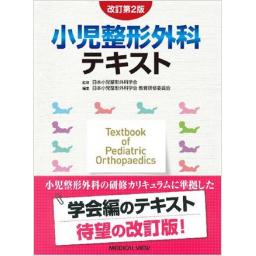
改訂第2版 小児整形外科テキスト

出版社: メジカルビュー社
著者:
発行日: 2016-12-10
分野: 臨床医学:外科  >  小児外科学
ISBN: 9784758313742
電子書籍版: 2016-12-10 (第2版第1刷)
書籍・雑誌
≪全国送料無料でお届け≫
取寄せ目安:8~14営業日

10,450 円(税込)

電子書籍
章別単位での購入はできません
ブラウザ、アプリ閲覧

10,450 円(税込)

商品紹介

改訂第2版では,日本小児整形外科学会 教育研修委員会のカリキュラムを基に,小児整形外科医を目指す医師に必要なすべての情報を網羅している。具体的には,診察法などの総論から始まり,検査,外傷,上肢疾患,下肢疾患,体幹,骨系統疾患,骨腫瘍,血液疾患,症候群,筋・神経疾患,炎症性疾患の12の章に分け,豊富な写真とイラストで,疾患の疫学から分類,治療法や予後について,丁寧に解説している。さらに,小児特有の事象は「Point」として強調することで,何が重要なのか一目でわかるようになっている。また,充実した付録が追加され,日常診療に則役立つ1冊となっている。

目次

  • 改訂第2版 小児整形外科テキスト

    ―目次―

    1 総論
    2 検査
    3 外傷
    4 上肢疾患
    5 下肢疾患
    6 体幹
    7 骨系統疾患
    8 骨腫瘍
    9 血液疾患
    10 症候群
    11 筋・神経疾患
    12 炎症性疾患

この書籍の参考文献

参考文献のリンクは、リンク先の都合等により正しく表示されない場合がありますので、あらかじめご了承下さい。

本参考文献は電子書籍掲載内容を元にしております。

I 総論

P.9 掲載の参考文献
1) 篠原寛休. 集団の性腺被曝を考慮した新しい乳児先天股脱検診の在り方について. 臨整外 1974;9:203-11.
2) 古森元章, ほか. 集団検診における幼児下肢軸の検討. 整外と災外 1986;35:684-7.
3) 和田 研, ほか. 幼児外反扁平足に対するcontrol study. 日足外科会誌 1989;10:103-6.
4) 高嶋明彦, ほか. 小児における生理的O脚の検討. 日小児整外会誌 1996;5:411-6.
5) 藤井敏男編. 小児の診察. 小児整形外科の実際. 東京:南山堂;2008. p1-6.
6) 藤井敏男編. 小児の診察のコツ. 整形外科Knack & Pitfalls 小児整形外科の要点と盲点. 東京:文光堂;2009. p2-13.
7) 藤井敏男, ほか編. 小児における疼痛・運動障害のみかた. 小児運動器疾患のプライマリケア 愁訴・症状からのアプローチ. 東京:南江堂;2015. p2-7.
8) 川浪 喬. X線上異常にみえやすいnormal variants. 小児運動器疾患のプライマリケア 愁訴・症状からのアプローチ. 藤井敏男, ほか編. 東京:南江堂;2015. p59-72
P.13 掲載の参考文献
1) Staheli LT, et al. Lower-extremity rotational problems in children. Normal values to guide management. J Bone Joint Surg Am 1985;67:39-47.
2) 町田治郎. 低リン血症性くる病. 今日の整形外科治療指針. 第7版. 土屋弘行, ほか編. 東京:医学書院;2016. p259-60.
3) 町田治郎. 先天性内転足. 最新整形外科学大系 18 下腿・足関節・足部. 越智光夫, ほか編. 東京:中山書店;2007. p130-4.
4) Jakob RP, et al. Tibial torsion calculated by computerized tomography and compared to other methods of measurement. J Bone Joint Surg Br 1980;62:238-42.
5) 小林秀男. CTによる下肢アライメント計測. 運動器の計測線・計測値ハンドブック. 紺野愼一編. 東京:南江堂;2012. p367-9.
6) Madadi F, et al. A new method for tibial torsion measurement by computerized tomography. J Orthop 2016;13:43-7.
7) 田中邦彦, ほか. 膝蓋大腿関節障害の検討 下肢回旋アライメントからみた新しい分類. 東京膝関節会誌 1989;10:44-51.
8) 稲葉 裕, ほか. O脚(2) 保存療法. 整形外科Knack & Pitfalls 小児整形外科の要点と盲点. 藤井敏男編. 東京:文光堂;2009. p182-3.
9) 松尾 隆. 脳性麻痺の下肢変形の治療. 日整会誌 1989;63:1426-35.
10) 町田治郎. 二分脊椎による足部変形に対する治療. 整形外科 Knack & Pitfalls 足の外科の要点と盲点. 山本晴康編. 東京:文光堂;2006. p307-13.
11) Herring JA, author. Tibial torsion. Tachdjian's Pediatric Orthopaedics. 5th ed. Philadelphia:Elsevier Saunders;2014. p739-41.
12) 松田蓉子, ほか. 大腿骨前捻角症候群における大腿骨減捻骨切り術に対する髄内釘の使用経験. 日小児整外会誌 2014;23:322-7.
P.19 掲載の参考文献
1) 横井広道. 一過性下肢痛(いわゆる成長痛). 小児科臨床 2013;66:2439-44.
2) 村上寶久. 不定期に反復する一過性下肢痛. 小児科 1991;32:1553-60.
3) 廣島和夫. 成長痛. 関節外科 1987;6:365-70.
4) Evans AM. Growing pains:contemporary knowledge and recommended practice. J Foot Ankle Res 2008;1:4-8.
5) Evans AM, et al. Prevalence of "growing pains" in young children. J Pediatr 2004;145:255-8.
6) 杉本義久, ほか. いわゆる成長痛について. 日小児整外会誌 1996;6:95-9.
7) 赤木龍一郎. 成長痛. こどもの整形外科疾患の診かた 診断・治療から患者家族への説明まで. 亀ヶ谷真琴編. 東京:医学書院;2011. p179-82.
8) Asadi-Pooya AA, et al. Are laboratory tests necessary in making the diagnosis of limb pains typical for growing pains in children? Pediatr Int 2007;49:833-5.
9) 横井広道. いわゆる成長痛(小児の一過性下肢痛)の診断基準作成の試み. 中四整外会誌 2014;26:65-8.
10) Sinigaglia R, et al. Musculoskeletal manifestations in pediatric acute leukemia. J Pediatr Orthop 2008;28:20-8.
11) 宮前多佳子. 小児の線維筋痛症. 臨床リウマチ 2014;26:266-74.
12) 井上雄一. レストレスレッグス症候群の臨床症状と診断. レストレスレッグス症候群(RLS) だからどうしても脚を動かしたい. 井上雄一, ほか編, 東京:アルタ出版;2009. p65-97.
13) Baxter MP, et al. "Growing pains" in childhood-a proposal for treatment. J Pediatr Orthop 1988;8:402-6.
14) 長野県立こども病院. みんな成長痛って知ってる? [http://nagano-child.jp/wp-content/themes/iryou_d2_tw/documents/iryou/seikeigeka/seichotuu.pdf].
15) Uziel Y, et al. Five-year outcome of children with "growing pains":correlations with pain threshold. J Pediatr 2010;156:838-40.
P.27 掲載の参考文献
1) Osgood RB. Lesions of the tibial tubercle occurring during adolescence. Boston Med Surg J 1903;148:114-7.
2) 大井淑雄. 骨端症. 整形外科および外傷学. 第1版. 森崎直木編. 東京:文光堂;1975. p203.
3) 芳賀信彦. 骨端症. Med Pract 2002;19:1068-70.
4) 熊澤雅樹, ほか. 当施設における踵骨骨端症の競技種目別特性. スポーツ医・科 2014;24:1-4.
5) Panner HJ. A peculiar affection of the capitulum humeri, resembling Calve-Perthes’ disease of the hip. Acta Radiol 1929;10:234-42.
6) Herring JA, author. Tachdjian’s Pediatric Orthopedics. 3rd ed. Philadelphia:Saunders;2002. p2207-8.
7) 鈴江直人, ほか. スポーツ選手における骨盤傷害. MB Orthop 2010;23(9):12-9.
8) 内田 理, ほか. ヴァン・ネック病. MB Orthop 1994;7(3):83-9.
P.32 掲載の参考文献
1) 藤井敏男. 小児整形外科の歴史. 臨整外 2015;50:1118-23.
2) 公益財団法人母子衛生研究会編集協力. 母子保健の主なる統計 平成27年度刊行. 東京:母子保健事業団;2016.
3) 公益財団法人母子衛生研究会編集協力. 母子保健の主なる統計 平成26年度刊行. 東京:母子保健事業団;2015.
P.35 掲載の参考文献
1) 徳村光昭. 学校健康診断における運動器検診マニュアル(2016年度実施案) 整形外科を専門としない学校医による実施方法. 慶應義塾大学保健管理センター;2016. [http://www.hcc.keio.ac.jp/japanese/education/locomotorium/2016.pdf]
P.45 掲載の参考文献
1) Whitby LG. Screening for disease:definitions and criteria. Lancet 1974;2:819-22.
2) 木下訓光, ほか. アスリートのメディカルチェックおよびその結果としての競技参加制限・中止勧告における社会的・法的・倫理的問題. 慶應義塾大学スポーツ医学研究センター紀要 1999;15-23.
3) 森原 徹, ほか. 京都府における小学生の投球障害肩・肘に対する早期発見・治療の取り組み. 日整外スポーツ医会誌 2013;33:19-26.
4) 松浦哲也, ほか. 少年野球肘検診-障害の早期発見・早期治療と予防を目指して. 関節外科 2008;27:1089-95.
5) 船越忠直, ほか. 超音波を用いた少年野球肘検診-病院受診率向上の工夫. JOSKAS 2012;37:8-9.
6) 森原 徹, ほか. 高校野球選手における肩関節のHyper External Rotation Test陽性率とそのセルフチェック法の有用性. 日臨スポーツ医会誌 2015;23:20-4.
7) 森原 徹, ほか. 京都における青少年に対するスポーツ検診の現状と課題. 日臨スポーツ医会誌 2014;22:395-401.
8) 立入克敏, ほか. 地域における取り組みの現状-京都市小学校「大文字駅伝」大会事前運動器検診を中心に. 運動器リハ 2013;24:26-32.
9) 松浦哲也, ほか. 少年野球選手の肘関節痛発症に関する前向き調査-危険因子の検討とガイドラインの検証. 日整外スポーツ医会誌 2012;32:242-7.

II 検査

P.52 掲載の参考文献
1) Graf R. Classification of hip joint dysplasia by means of sonography. Arch Orthop Trauma Surg 1984;102:248-55.
2) Kramps HA, et al. Einsatzmoglichkeiten der Ultraschalldiagnostik am Bewegungsapparat. Z Orthop 1979;118:355-64.
3) Wilson DJ, et al. Arthro sonography of the painful hip. Clin Radiol 1984;35:17-9.
4) Kallio P, et al. Ultrasonography in hip disease in children. Acta Orthop Scand 1985;56:367-71.
P.57 掲載の参考文献
1) Kan JH, et al. Value of MRI after recent diagnostic or surgical intervention in children with suspected osteomyelitis. AJR Am J Roentgenol 2008;191:1595-600.
2) Mignemi ME, et al. Epidemiology, diagnosis, and treatment of pericapsular pyomyositis of the hip in children. J Pediatr Orthop 2014;34:316-25.
3) Nakamura J, et al. Hip arthrography under general anesthesia to refine the definition of hinge abduction in Legg-Calve-Perthes disease. J Pediatr Orthop 2008;28:614-8.
4) Kamegaya M, et al. Arthrographic indicators for decision making about femoral varus osteotomy in Legg-Calve-Perthes disease. J Child Orthop 2008;2:261-7.

III 外傷

P.74 掲載の参考文献
1) Rockwood CA, et al, editors. Fractures in children. New York:Lippincott-Raven Press;1996.
2) Rang M. Children's fracture. 2nd ed. Philadelphia:Lippincott;1984.
3) 河本浩栄, ほか. 小児骨折に対する骨端線損傷の疫学的検討. 日小児整外会誌 1999;8:196-200.
4) 河本浩栄, ほか. 開業医院での小児骨端線損傷の疫学調査. 日小児整外会誌 2000;9:259-63.
5) 廣島和夫. 整形外科からみた児童虐待. 医療 2012;66:305-10.
6) Heffernan MJ, et al. Treatment of Femur Fractures in Young Children:A Multicenter Comparison of Flexible Intramedullary Nails to Spica Casting in Young Children Aged 2 to 6 Years. J Pediatr Orthop 2015;35:126-9.
P.79 掲載の参考文献
1) 麻生邦一. 肘内障の臨床的研究-とくに受傷機転と治療法の検討-. 日小児整外会誌 2008;17:122-6.
2) 今村惠一郎, ほか. 小児肘内障の超音波診断. 関節外科 2012;31:404-11.
3) Jakob R, et al. Observations concerning fractures of the lateral humeral condyle in children. J Bone Joint Surg Br 1975;57:430-6.
4) 今田英明, ほか. 小児上腕骨外顆骨折の3次元的形態および上腕骨20°挙上位撮影法の有用性に関する検討. 骨折 2010;32:5-11.
5) Zhang JD, et al. Ultrasonography for non-displaced and minidisplaced humeral lateral condyle fractures in children. Chin J Traumatol 2008;11:297-300.
6) Kamegaya M, et al. Assessment of stability in children's minimally displaced lateral humeral condyle fracture by magnetic resonance imaging. J Pediatr Orthop 1999;19:570-2.
7) Song KS, et al. Closed reduction and internal fixation of displaced unstable lateral condylar fractures of the humerus in children. J Bone Joint Surg Am 2008;90:2673-81.
8) 洪 淑貴, ほか. 小児上腕骨外側顆骨折後内反肘変形の治療経験. 日肘関節会誌 2013;20:197-9.
9) 大島 明, ほか. 小児肘関節外側顆骨折の治療成績. 日肘関節会誌 2016;22:S107.
10) 川崎恵吉, ほか. 小児上腕骨外側顆骨折に対する手術成績の比較-Kirschner鋼線固定法とTension Band Wiring法-. 日肘関節会誌 2013;20:163-6.
11) Shimada K, et al. Osteosynthesis for the treatment of nonunion of the lateral humeral condyle in children. J Bone Joint Surg Am 1997;79:234-40.
12) Otsuka J, et al. Unusual humeral medial condyle fracture in an adolescent because of a previous post-traumatic fishtail deformity:a case report. J Pediatr Orthop B 2015;24:408-11.
P.91 掲載の参考文献
1) 佐々木 孝. 上腕骨顆上骨折に対する観血的治療法. MB Orthop 1993;6(4):63-70.
2) Rockwood CA, et al, Authors. Fractures in Children. 2nd ed. Philadelphia:J.B.Lippincott;1984. p383.
3) Yamamoto I, et al. Cubitus varus deformity following supracondylar fracture of the humerus. A method for measuring rotational deformity. Clin Ortop Relat Res 1985;201:179-85.
4) 稲垣克記, ほか. 小児上腕骨顆上骨折-教室過去400例の検討から-. 日小児整外会誌 2005;14:159-63.
5) 藤巻悦夫, ほか. 内反肘矯正骨切術の検討. 整・災外 1983;26:1597-604.
6) 伊藤恵康. 上腕骨顆上骨折. 骨折・脱臼. 改訂2版. 富士川恭輔, ほか著. 東京:南山堂;2005. p313.
7) 大口怜央, ほか. 小児上腕骨顆上骨折に対する経皮的ピンニング後に生じた神経障害の検討. 骨折 2009;31:371-4.
P.97 掲載の参考文献
1) 伊藤惠康著. Monteggia骨折. 肘関節外科の実際 私のアプローチ. 東京:南江堂;2011. p172-85.
2) 森久喜八郎. モンテジア損傷. 整外MOOK 1988;54:112-27.
4) 上村正吉, ほか. 小児の骨折・肘関節部の外傷. 整外MOOK 1980;13:110-31.
5) Borden S 4th. Traumatic bowing of the forearm in children. J Bone Joint Surg Am 1974;56:611-6.
6) Lincoln TL, et al. "Isolated"traumatic radial-head dislocation. J Pediatr Orthop 1994;14:454-7.
7) Letts M, et al. Monteggia fracture-dislocations in children. J Bone Joint Surg Br 1985;67:724-7.
8) Altner PC. Monteggia fractures. Orthop Review 1981;10:115-20.
9) Spinner M, et al. Posterior interosseous nerve palsy as a complication of Monteggia fractures in children. Clin Orthop Relat Res 1968;58:141-5.
10) 蔵本哲也, ほか. 腕橈関節に陥入していた後骨間神経を術中に確認したMonteggia骨折の1例. 日肘関節会誌 2007;14:S62.
P.102 掲載の参考文献
1) 斧出絵麻, ほか. 小児大腿骨頚部骨折における受傷機転の検討. 整形外科 2014;65:153-6.
2) Ratliff AH. Fractures of the neck of the femur in children. J Bone Joint Surg Br 1962;44:528-42.
3) Beaty JH. Fractures of the hip in children. Orthop Clin North Am 2006;37:223-32.
4) Delbet MP. Fractures du col de femur. Bull Mem Soc Chir 1909;35:387-9.
5) Colonna PC. Fracture of the neck of the femur in children. Am J Surg 1929;6:793-7.
6) 井上 博著. 大腿骨頚部骨折. 小児四肢骨折治療の実際. 第2版. 東京:金原出版;2001. p314-23.
7) 松田秀一, ほか. 大腿骨頚部骨折に対する整復と内固定法. OS Now Instruction No.1 小児の骨折・外傷. 岩本幸英, ほか編. 東京:メジカルビュー社;2007. p146-57.
8) 土屋正光, ほか. 小児大腿骨頚部骨折. 新OS Now No.4 小児外傷の保存療法と手術療法. 落合直之, ほか編. 東京:メジカルビュー社;1999. p90-7.
9) 北 純, ほか. 小児の大腿骨近位部骨折. 股関節学. 久保俊一編著. 京都:金芳堂;2014. p529-39.
10) Canale ST, et al. Pelvic and hip fractures. Fractures in Children. Rockwood CA Jr, et al, editors. Philadelphia:Lippincott Williams & Wilkins:1984. p733-843.
11) 岩崎勝郎. 大腿骨頚部骨折. OS Now No.10 関節周辺骨折の治療. 林 浩一郎, ほか編. 東京:メジカルビュー社;1993. p78-87.
12) Hedlund R, et al. The incidence of femoral shaft fractures in children and adolescents. J Pediatr Orthop 1986;6:47-50.
13) 井上 博著. 大腿骨骨幹部骨折. 小児四肢骨折治療の実際. 第2版. 東京:金原出版;2001. p335-54.
14) 佐藤栄一, ほか. 大腿骨骨幹部骨折に対する創外固定法. OS Now Instruction No.1 小児の骨折・外傷. 岩本幸英, ほか編. 東京:メジカルビュー社;2007. p158-68.
15) 前 隆男. 大腿骨骨幹部骨折に対する懸垂, 牽引療法と髄内釘法. OS Now Instruction No.1 小児の骨折・外傷. 岩本幸英, ほか編. 東京:メジカルビュー社;2007. p169-82.
16) Flynn JM, et al. Femoral shaft fractures. Rockwood and Wilkins' Fractures in Children. 8th ed. Flynn JM, et al, editors. Indianapolis:Wolters Kluwer;2014. p987-1026.
P.106 掲載の参考文献
1) 福林 徹. 平成21~23年度における3年間のまとめ. 日体育協会スポーツ医科研報集 2013;2012:48-53.
2) 奥脇 透. 平成21~23年度における3年間のまとめ. 日体育協会スポーツ医科研報集 2013;2012:10-22.
3) 鳥居 俊. こどもの身体の特徴(総論). こどものスポーツ障害診療ハンドブック. 山下敏彦編. 東京:中外医学社;2013. p1-7.
4) 戸祭正喜, ほか. プロサッカークラブの下部組織におけるスポーツ外傷および障害の発生状況. 関節外科 2008;27:1693-700.
P.113 掲載の参考文献
1) Kempe CH, et al. The battered-child syndrome. JAMA 1962;181:17-24.
2) King J, et al. Analysis of 429 fractures in 189 battered children. J Pediatric Orthop 1988;8:585-9.
3) Kleinman PK, et al. The metaphyseal lesion in abused infants:a radiologic-histopathologic study. Am J Roentgenol 1986;146:895-905.
4) Kleinman PK. Diagnotic imaging of child abuse. 2nd ed. St. Louis:Mosby;1998.

IV 上肢疾患

P.123 掲載の参考文献
1) Birch R, author. Birth lesion of the brachial plexus. Surgical disorders of the peripheral nerves. Philadelphia:Churchill Livingstone;2011. p429-82.
2) 名倉温雄, ほか. 分娩麻痺の予後と合併分娩外傷との関係について. 大阪母子保健総合医療セ誌 2004;20:20.
3) Foad SL, et al. The epidemiology of neonatal brachial plexus palsy in the United States. J Bone Joint Surg Am 2008;90:1258-64.
4) Wall LB, et al. Incidence and prognosis of neonatal brachial plexus palsy with and without clavicle fractures. Obstet Gynecol 2014;123:1288-93.
5) 川端秀彦, ほか. 分娩麻痺の作業療法・運動療法. 運動療物理療 2007;18:2-7.
6) 川端秀彦. 分娩麻痺の治療戦略-上位型麻痺における神経修復術の適応について. 日整会誌 2013;87:43-7.
7) Waters PM. Update on management of pediatric brachial plexus palsy. J Pediatr Orthop A 2005;25:116-26.
8) 川端秀彦. 分娩麻痺. 最新整形外科学大系 15A 手関節・手指I. 三浪明男, ほか編. 東京:中山書店;2007. p166-75.
9) 川端秀彦. 新生児・乳児に見られる骨折. MB Orthop 2002;15(12):16-23.
10) Madsen ET. Fractures of the extremities in the newborn. Acta Obstet Gynecol Scand 1955;34:41-74.
11) 藤井敏男, ほか. 分娩骨折の診断, 治療上の問題点と予後. 整・災外 1990;33:5-12.
P.130 掲載の参考文献
1) Swanson AB. A classification for congenital limb malformations. J Hand Surg Am 1976;1:8-22.
2) Iba K, et al. The Classifi cation of Swanson for congenital 2 anomalies of upper limb modified by the Japanese society for surgery of the hand 3(JSSH). Hand Surg 2015;20:237-50.
3) Tonkin MA, et al. Classification of congenital anomalies of the hand and upper limb:development and assessment of a new system. J Hand Surg Am 2013;38:1845-53.
4) Wassel HD. The results of surgery for polydactyly of the thumb. A review. Clin Orthop Relat Res 1969;64:175-93.
5) Blauth W. Der hypoplastische Daumen. Arch Orthop Unfall-Chirur 1967;62:225-46.
6) Bayne LG, et al. Long-term review of the surgical treatment of radial deficiencies. J Hand Surg Am 1987;12:169-79.
7) Takayama S, et al. Modified abductor digiti minimi opponensplasty in congenital hypoplastic thumb with laxity of metacarpophalangeal joint. Tech Hand Up Extrem Surg 2002;6:166-70.
8) 高山真一郎, ほか. 母指形成不全. PEPARS 2015;103:48-58.

V 下肢疾患

P.139 掲載の参考文献
1) 品田良之, ほか. 松戸市の乳児先天性股関節脱臼検診の現状と今後. 整形外科 2014;65:1017-22.
2) 和田郁雄, ほか. リーメンビューゲル(Rb)治療マニュアル-先天性股関節脱臼(発育性股関節形成不全)に対する安全な装着を目指して-日本小児股関節研究会リーメンビューゲル治療に関するワーキンググループ作成 平成23年度版Ver1.2. 日小児整外会誌 2012;21:391-408.
3) Yamamuro T, et al. A radiological study on the development of the hip joint in normal infants. J Jpn Orthop Assoc 1975;49:421-39.
5) Graf R. Classification of hip joint dysplasia by means of sonography. Arch Orthop Trauma Surg 1984;102:248-55.
6) 池田 威, ほか. 先天性股関節脱臼の保存的療法 リーメンビューゲル法. 整・災外 1998;41:409-15.
7) 和田郁雄, ほか. 我が国での先天股脱に対するリーメンビューゲル治療の現状. 日小児整外会誌 2009;18:272-5.
P.149 掲載の参考文献
1) Salter RB, et al. Legg-Calve-Perthes disease. The prognostic significance of the subchondral fracture and a two-group classification of the femoral head involvement. J Bone Joint Surg Am 1984;66:479-89.
2) Catterall A. The natural history of Perthes' disease. J Bone Joint Surg Br 1971;53:37-53.
3) Herring JA, et al. Legg-Calve-Perthes disease. Part I:Classification of radiographs with use of the modified lateral pillar and Stulberg classifications. J Bone Joint Surg Am 2004;86:2103-20.
4) Staheli LT, author. Legg-Calve-Perthes disease. Fundamentals of Pediatric Orthopedics. 4th ed. Philadelphia:Lippincott Williams & Wilkins;2008. p218-23.
5) Herring JA, et al. The lateral pillar classification of Legg-Calve-Perthes Disease. J Pediatr Orthop 1992;12:143-50.
6) Mose K. Methods of measuring in Legg-Calve-Perthes disease with special regard to the prognosis. Clin Orthop Relat Res 1980;150:103-9.
7) Stulberg SD, et al. The natural history of Legg-Calve-Perthes Disease. J Bone Joint Surg Am 1981;63:1095-108.
P.158 掲載の参考文献
1) Kay RM. Slipped Capital Femoral Epiphysis. Lovell and Winter's Pediatric Orthopaedics. 6th ed. Morrissy RT, et al, editors. Philadelphia:Lippincott Wlliams & Wilkins;2005. p1085-124.
2) Lehmann CL, et al. The epidemiology of slipped capital femoral epiphysis:an update. J Pediatr Orthop 2006;26:286-90.
3) Noguchi Y, et al. Epidemiology and demographics of slipped capital femoral epiphysis in Japan:a multicenter study by the Japanese Paediatric Orthopaedic Association. J Orthop Sci 2002;7:610-7.
4) Loder RT, et al. Acute slipped capital femoral epiphysis:the importance of physeal stability. J Bone Joint Surg Am 1993;75:1134-40.
5) Guzzanti V, et al. Slipped capital femoral epiphysis:comparison of a roentgenographic method and computed tomography in determining slip severity. J Pediatr Orthop 1991;11:6-12.
6) Jerre R, et al. Bilaterality in slipped capital femoral epiphysis:importance of a reliable radiographic method. J Pediatr Orthop B 1996;5:80-4.
7) Clohisy JC, et al. A systematic approach to the plain radiographic evaluation of the young adult hip. J Bone Joint Surg Am 2008;90 Suppl 4:47-66.
8) Roaten J, et al. Complications related to the treatment of slipped capital femoral epiphysis. Orhtop Clin North Am 2016;47:405-13.
P.163 掲載の参考文献
1) Charif P, et al. Genu recurvatum congenitum in the newborn:its incidence, course, treatment, prognosis. Clin Pediatr(Phila) 1965;4:587-94.
2) Jacobsen K, et al. Congenital dislocation of the knee. Acta Orthop Scand 1985;56:1-7.
3) Bensahel H, et al. Congenital dislocation of the knee. J Pediatr Orthop 1989;9:174-7.
4) Leveuf J, et al. Les dislocations congenitales du genou(genu recurvatum, subluxation, luxation). Rev Chir Orthop 1947;32:313-50.
5) Haga N, et al. Congenital dislocation of the knee reduced spontaneously or with minimal treatment. J Pediatr Orthop 1997;17:59-62.
6) Roy DR, et al. Percutaneous quadriceps recession:a technique for management of congenital hyperextension deformities of the knee in the neonate. J Pediatr Orthop 1989;9:717-9.
7) Wilson RL, et al. Congenital hyperextension of the knee. Tachdjian's pediatric orthopaedics. 5th ed. Herring JA, editor. Philadelphia:Elsevier;2014. p682-6.
8) Wada A, et al. Surgical treatment of hip dislocation in amyoplasia-type arthrogryposis. J Pediatr Orthop B 2012;21:381-5.
P.171 掲載の参考文献
1) Salenius P, et al. The development of the tibiofemoral angle in children. J Bone Joint Surg Am 1975;57:259-61.
2) Blount WP. Tibia var, osteochondrosis deformans tibiae. J Bone Joint Surg 1937;19:1-29.
3) Langenskiold A, et al. Tibia vara(osteochondrosis deformans tibiae):A survey of seventy-one cases. J Bone Joint Surg Am 1964;46:1405-20.
4) Inaba Y, et al. Multicenter study of Blount disease in Japan by the Japanese Pediatric Orthopaedic Association. J Orthop Sci 2014;19:132-40.
5) Levine AM, et al. Physiological bowing and tibia vara. The metaphyseal-diaphyseal angle in the measurement of bowleg deformities. J Bone Joint Surg Am 1982;64:1158-63.
6) Zayer M. Long-term results after physiological genu varum. J Pediatr Orthop B 2000;9:271-7.
P.176 掲載の参考文献
1) Crawford AH, et al. Neurofibromatosis in children:the role of the orthopaedist. J Am Acad Orthop Surg 1999;7:217-30.
2) Hefti F, et al. Congenital pseudarthrosis of the tibia:history, etiology, classification, and epidemiologic data. J Pediatr Orthop B 2000;9:11-5.
3) Boyd HB. Pathology and natural history of congenital pseudarthrosis of the tibia. Clin Orthop Relat Res 1982;166:5-13.
4) Crawf ord AH Jr, et al. Osseous manifestations of neurofibromatosis in childhood. J Pediatr Orthop 1986;6:72-88.
5) Crawford AH. Neurofibromatosis in children. Acta Orthop Scand Suppl 1986;218:1-60.
6) Herring JA, author. Tachdjian's Pediatric Orthopaedics. 5th ed. Philadelphia:Elsevier;2014.
7) Kitoh H, et al. Congenital anterolateral bowing of the tibia with ipsilateral polydactyly of the great toe. Am J Med Genet 1997;73:404-7.
8) Manner HM, et al. Pathomorphology and treatment of congenital anterolateral bowing of the tibia associated with duplication of the hallux. J Bone Joint Surg Br 2005;87:226-30.
P.182 掲載の参考文献
1) Yamamoto H. A clinical, genetic and epidemiologic study of congenital club foot. Jap J Human Genet 1979;24:37-44.
2) Ponseti I IV, author. Congenital Clubfoot:Fundamentals of Treatment. Oxford:Oxford University Press;1996.
3) Staheli L. 先天性内反足:Ponseti法. 第3版. 日本語版. Seattle:Global-Help organization:2010. [http://www.global-help.org/publications/books/help_cfponsetijapanese.pdf].
4) 北 純. 先天性内反足. 整形外科 2012;63:673-81.
5) 北 純. 先天性内反足. 整形外科臨床パサージュ 9. 足の痛み クリニカルプラクティス. 中村耕三, 木下光雄編. 東京:中山書店;2011. p306-15.
6) Dimeglio A. Classification of clubfoot. J Pediatr Orthop B 1995;4:129-36.
7) Pirani S. A method of assessing the virgin clubfoot. Orlando:Pediatric Orthopaedic Society of North America(POSNA);1995.
8) 山本晴康. 後内側解離術. 小児整形外科 手術テクニック. 日本小児整形外科学会教育研修委員会編. 東京:メジカルビュー社;2007. p62-73.
9) 北 純. 距骨下関節全周解離術. 小児整形外科 手術テクニック. 日本小児整形外科学会教育研修委員会編. 東京:メジカルビュー社;2007. p74-86.
P.188 掲載の参考文献
1) Lichtblau S. Congenital vertical talus. Bull Hosp Joint Dis 1978;39:165-79.
2) Wynne-Davis R. Genetics and congenital musculoskeletal disorders. Obstet Gynecol Annu 1977;6:247-60.
3) Kite JH. Congenital metatarsus varus. J Bone Join Joint Surg Am 1967;49:388-97.
4) Herring JA, author. Tachdjian's Pediatric Orthopaedics Vol 2. 5th Ed. Philadelphia:Saunders;2013.
P.194 掲載の参考文献
1) Stormont DM, et al. The relative incidence of tarsal coalition. Clin Orthop Relat Res 1983;181:28-36.
2) Harris RH, et al. Hypermobile flat-foot with short tendo Achillis. J Bone joint Surg Am 1948;30:116-40.
3) Bleck EE, et al. Conservative management of pes valgus with plantar flexed talus, flexible. Clin Orthop Relat Res 1977;122:85-94.
4) Bordelon RL. Correction of hypermobile flatfoot in children by molded insert. Foot Ankle 1980;1:143-50.
5) 和田郁雄, ほか. 小児外反扁平足の病態と治療. 整・災外 2004;47:1131-9.
6) Moraleda L, et al. Comparison of the calcaneo-cuboidcuneiform osteotomies and the calcaneal lengthening osteotomy in the surgical treatment of symptomatic flexible flatfoot. J Pediatr Orthop 2012;32:821-9.
7) 薩摩眞一. こどもの扁平足:硬い扁平足. MB Orthop 2013;26(6):25-32.
P.205 掲載の参考文献
1) Frantz CH, O'Rahilly R. Congenital Skeletal Limb Deficiencies. J Bone Joint Surg Am 1961;43:1202-24.
2) 廣島和夫. 2 下肢の異常. 小児整形外科テキスト. 日本小児整形外科学会教育研修委員会編. 東京:メジカルビュー社;2004. p29-36.
3) Lowry RB, Bedard T. Congenital limb deficiency classification and nomenclature:The need for a consensus. Am J Med Genet A 2016;170:1400-4.
4) Swanson AB. A classification for congenital malformations of the hand. Acad Med Bull New Jersey 1964;10:166-9.
5) Kay H. A proposed international terminology for the classification of congenital limb deficiencies. Orthotics Prosthetics 1974;28:33-48.
6) Stoll C, Mastroiacovo P, de Wals P, et al. EUROCAT Guide 3:For the description and classification of congenital limb defects. 2nd ed. Catholic University of Louvain. Belgium:EUROCAT Central Registry;1986.
7) Ogino T. Congenital anomalies of the hand. The Asian perspective. Clin Orthop Relat Res 1996;323:12-21.
8) Oberg KC, Feenstra JM, Manske PR, et al. Developmental biology and classification of congenital anomalies of the hand and upper extremity. J Hand Surg Am 2010;35:2066-76.
9) Tonkin MA, Tolerton Sk, Quick TJ, et al. Classification of congenital anomalies of the hand and upper limb:development and assessment of a new system. J Hand Surg Am 2013;38:1845-53.
10) Gillbert SF著, 阿形清和, 高橋淑子監訳. ギルバート発生生物学. 東京:メディカルサイエンスインターナショナル;2015.
11) Al-Qattan MM, Kozin SH. Update on embryology of the upper limb. J Hand Surg Am 2013;38:1835-44.
12) Nowack E. Die sensible phase bei der thalidomidembryopathie. Humangenetik 1965;1:516-36.
13) Pappas AM. Congenital abnormalities of the femur and related lower extremity malformations:classification and treatment. J Pediatr Orthop 1983;3:45-60.
14) Syvanen J, Nietosvaara Y, Ritvanen A, et al. High risk for major nonlimb anomalies associated with lower-limb deficiency:a population-based study. J Bone Joint Surg Am 2014;96:1898-904.
15) Kalamchi A, Dawe RV. Congenital deficiency of the tibia. J Bone Joint Surg Br 1985;67:581-4.
16) Achterman C, Kalamchi A. Congenital deficiency of the fibula. J Bone Joint Surg Br 1979;61:133-7.
17) Boyd HB. Pathology and natural history of congenital pseudarthrosis of the tibia. Clin Orthop Relat Res 1982;166:5-13.
P.214 掲載の参考文献
1) 高橋光彦. 下肢長不等をきたす疾患とその対処法. 小児整形外科テキスト. 日本小児整形外科学会教育研修委員会編. 東京:メジカルビュー社;2004. p196-202.
2) 吉田泰久, ほか. 脚長差による機能的臼蓋形成不全に対し脚延長で治療を行った2例. 日本創外固定・骨延長学会雑誌 2016;27:41-5.
3) 渡部欣忍, ほか. トモシンセシスとdigital slit scanogramの原理と運動器外傷領域への活用. 整形外科 2016;67:462-8.
4) Moseley CF, et al. A straight-line graph for leg-length discrepancies. J Bone Joint Surg Am 1977;59:174-9.
5) Paley D, et al. Multiplier method for predicting limb-length discrepancy. J Bone Joint Surg Am 2000;82:1432-46.
6) Sabharwal S, et al. Growth patterns after lengthening of congenitally short lower limbs in young children. J Pediatr Orthop 2000;20:137-45.
7) Song KM, et al. The effect of limb-length discrepancy on gait. J Bone Joint Surg Am 1997;79:1690-8.
8) 田村太資. 脚長差に伴う跛行とその対策. MB Orthop 2015;28(4):27-32.
9) 滝川一晴, ほか. 脚長不等に対する経皮的膝骨端線閉鎖術の治療効果. 日小児整外会誌 2006;15:50-4.
10) Metaizeau JP, et al. Percutaneous epiphysiodesis using transphyseal screws(PETS). J Pediatr Orthop 1998;18:363-9.
11) Pendleton AM, et al. Guided growth for the treatment of moderate leg-length discrepancy. Orthopedics 2013;36:e575-80.
12) 森川耀源, ほか. エイトプレートを用いた骨端線成長抑制術の神経線維腫症1型に対する有効性. 日小児整外会誌 2015;24:48-52.
13) 櫻吉啓介, ほか. Postaxial hypoplasiaに対する下肢延長術の治療成績と問題点. 日小児整外会誌 2005;14:71-6.
14) Paley D. Problems, obstacles, and complications of limb lengthening by the Ilizarov technique. Clin Orthop Relat Res 1990;250:81-104.

VI 体幹

P.221 掲載の参考文献
1) Copley LAB. Congenital Muscular Torticollis. Tachdjian's Pediatric Orthopaedics. 5th ed. Herring JA, author. Philadelphia:Saunders;2014. p167-73.
2) 篠田達明, ほか. 乳児筋性斜頸のいわゆる自然治ゆについて. 臨整外 1970;5:82-8.
3) 篠田達明. 乳児筋性斜頸マッサージに対する疑問. 整形外科 1971;22:22-9.
4) Akazawa H, et al. Congenital muscular torticollis:longterm follow-up of thirty-eight partial resections of the sternocleidomastoid muscle. Arch Orthop Trauma Surg 1993;112:205-9.
5) 中塚洋一, ほか. 就学年令以後に行った筋性斜頸の手術成績. 中部整災誌 1986;29:1355-65.
6) 田辺剛造. 先天性筋性斜頚-観血的療法とその成績-. 日整会誌 1981;55:807-17.
7) Fielding JW, et al. Atlanto-axial rotatory fixation. (Fixed rotatory subluxation of the atlanto-axial joint). J Bone and Joint Surg Am;59:37-44.
P.227 掲載の参考文献
1) Purkiss SB, et al. Idiopathic scoliosis in families of children with congenital scoliosis. Clin Orthop Relat Res 2002;401:27-31.
2) Winter RB. Congenital scoliosis. Clin Orthop Relat Res 1973;93:75-94.
3) Shands AR Jr, et al. The incidence of scoliosis in the state of Delaware;a study of 50,000 minifilms of the chest made during a survey for tuberculosis. J Bone Joint Surg Am 1955;37:1243-9.
4) Giampietro PF, et al. Congenital and idiopathic scoliosis:clinical and genetic aspects. Clin Med Res 2003;1:125-36.
5) Belmont PJ Jr, et al. Intraspinal anomalies associated with isolated congenital hemivertebra:the role of routine magnetic resonance imaging. J Bone Joint Surg Am 2004;86:1704-10.
6) 駒形正志. 先天性脊柱側弯症. 今日の整形外科治療指針. 第7版. 土屋弘行, ほか編. 東京:医学書院;2016. p585-6.
7) Hedequist D, et al. Congenital scoliosis. J Am Acad Orthop Surg 2004;12:266-75.
8) Hedequist D, et al. Congenital scoliosis. a review and update. J Pediatr Orthop 2007;27:106-16.
9) Kawakami N, et al. Classification of congenital scoliosis and kyphosis:a new approach to the three-dimensional classification for progressive vertebral anomalies requiring operative treatment. Spine(Phila Pa 1976) 2009;34:1756-65.
10) McMaster MJ, et al. The Natural History of congenital scoliosis. A study of two hundred and fi fty-one patients. J Bone Joint Surg Am 1982;64:1128-47.
P.231 掲載の参考文献
1) Reisenborough EJ, et al. A genetic survey of idiopathic scoliosis in Boston, Massachusetts. J Bone Joint Surg Am 1973;55:974-82.
2) Lonstein JE, et al, authors. Idiopathic scoliosis. Moe's textbook of scoliosis and other spinal deformities. 3rd ed. Philadelphia:W.B. Saunders;1995. p219-56.
3) 瀬本喜啓. 装具治療. 側弯症治療の最前線-基礎編. 日本側彎症学会編. 大阪:医薬ジャーナル社;2013. p136.
P.238 掲載の参考文献
1) Madigan RR, et al. Scoliosis in the institutionalized cerebral palsy population. Spine(Phila Pa 1976) 1981;6:583-90.
2) Majd ME, et al. Natural history of scoliosis in the institutionalized adult cerebral palsy population. Spine(Phila Pa 1976) 1997;22:1461-6.
3) Lonstein JE, et al. Operative treatment of spinal deformities in patients with cerebral palsy or mental retardation. An analysis of one hundred and seven cases. J Bone Joint Surg Am 1983;65:43-55.
4) Nakamura N, et al. Use of dynamic spinal brace in the management of neuromuscular scoliosis:a preliminary report. J Pediatr Orthop B 2014;23:291-8.
5) 吉田 清, ほか. 脳性麻痺の脊柱側弯変形に対する動的脊柱装具の介護者からみた効果の検討. The Japanese Journal of Rehabilitation Medicine 2015;52:251-5.
6) McMaster WC, et al. Spinal bracing in the institutionalized person with scoliosis. Spine(Phila Pa 1976) 1980;5:459-2.
7) Nash CL, Jr. Current concepts review:scoliosis bracing. J Bone Joint Surg Am 1980;62:848-52.
8) Canavese F, et al. Surgical advances in the treatment of neuromuscular scoliosis. World J Orthop 2014;5:124-33.
9) Sarwark J, et al. New strategies and decision making in the management of neuromuscular scoliosis. Orthop Clin North Am 2007;38:485-96.
10) Teli MG, et al. Spinal fusion with Cotrel-Dubousset instrumentation for neuropathic scoliosis in patients with cerebral palsy. Spine(Phila Pa 1976) 2006;31:E441-7.
11) Tsirikos AI, et al. Comparison of parents' and caregivers' satisfaction after spinal fusion in children with cerebral palsy. J Pediatr Orthop 2004;24:54-8.
12) Watanabe K, et al. Is spine deformity surgery in patients with spastic cerebral palsy truly beneficial?:a patient/parent evaluation. Spine(Phila Pa 1976) 2009;34:2222-32.
13) Saito N, et al. Natural history of scoliosis in spastic cerebral palsy. Lancet 1998;351:1687-92.
14) Fienman NL. Pediatric neurofibromatosis:review. Compr Ther 1981;7:66-72.
15) Neurofibromatosis. Conference statement. National Institutes of Health Consensus Development Conference. Arch Neurol 1988;45:575-8.
16) Winter RB, et al. Spine deformity in neurofibromatosis. A review of one hundred and two patients. J Bone Joint Surg Am 1979;61:677-94.
17) Calvert PT, et al. Scoliosis in neurofibromatosis. The natural history with and without operation. J Bone Joint Surg Br 1989;71:246-51.
18) Hsu LC, et al. Dystrophic spinal deformities in neurofibromatosis. Treatment by anterior and posterior fusion. J Bone Joint Surg Br 1984;66:495-9.
19) Loeys BL, et al. The revised Ghent nosology for the Marfan syndrome. J Med Genet 2010;47:476-85.
20) Sponseller PD, et al. Results of brace treatment of scoliosis in Marfan syndrome. Spine(Phila Pa 1976) 2000;25:2350-4.
21) Sponseller PD, et al. Infantile scoliosis in Marfan syndrome. Spine(Phila Pa 1976) 1997;22:509-16.
22) Lipton GE, et al. Surgical treatment of scoliosis in Marfan syndrome:guidelines for a successful outcome. J Pediatr Orthop 2002;22:302-7.
23) Dobbs MB, et al. Prevalence of neural axis abnormalities in patients with infantile idiopathic scoliosis. J Bone Joint Surg Am 2002;84:2230-4.
24) 徳永 誠, ほか. 側彎症外来初診患者におけるMRI所見の検討. 脊柱変形 2000;15:7-11.
25) Charry O, et al. Syringomyelia and scoliosis:a review of twenty-five pediatric patients. J Pediatr Orthop 1994;14:309-17.
26) Phillips WA, et al. Management of scoliosis due to syringomyelia in childhood and adolescence. J Pediatr Orthop 1990;10:351-4.
27) Eule JM, et al. Chiari I malformation associated with syringomyelia and scoliosis:a twenty-year review of surgical and nonsurgical treatment in a pediatric population. Spine(Phila Pa 1976) 2002;27:1451-5.
28) Yeom JS, et al. Scoliosis associated with syringomyelia:analysis of MRI and curve progression. Eur Spine J 2007;16:1629-35.
29) Sha S, et al. Evolution of syrinx in patients undergoing posterior correction for scoliosis associated with syringomyelia. Eur Spine J 2015;24:955-62.
30) Wang G, et al. One-Stage Correction Surgery of Scoliosis Associated With Syringomyelia:Is it Safe to Leave Untreated a Syrinx Without Neurological Symptom? J Spinal Disord Tech 2015;28:E260-4.
P.243 掲載の参考文献
1) 加藤真介, ほか. 発育期の後方終板障害と椎間板ヘルニア. MB Orthop 2003;16(9):42-7.
2) 加藤真介, ほか. 発育期におけるスポ-ツと腰痛 腰椎分離症と終板障害の病態と治療. 脊椎脊髄ジャーナル 2000;13:496-506.
3) Jackson DW, et al. Spondylolysis in the female gymnast. Clin Orthop Relat Res 1976;117:68-73.
4) 松本 學. 腰部障害の保存的治療. 臨スポーツ医 2006;23:1301-9.
5) Seitsalo S, et al. Spondylolistesis in ballet dancers. J Dance Med Sci 1997;1:51-4.
6) Soler T, et al. The prevalence of spondylolysis in the Spanish elite athlete. Am J Sports Med 2000;28:57-62.
7) Sairyo K, et al. MRI signal changes of the pedicle as an indicator for early diagnosis of spondylolysis in children and adolescents:a clinical and biomechanical study. Spine(Phila Pa 1976) 2006;31:206-11.
8) 西良浩一. 発育期腰椎分離症の早期診断と保存療法の最前線つくかつかないか, すべるかすべらないか. J Spine Res 2010;1:30-40.
9) 竹林庸雄. 腰椎終板障害. スポーツと腰痛 メカニズム&マネジメント. 山下敏彦編. 東京:金原出版;2011. p65-8.
10) 竹林庸雄, ほか. 腰椎椎間板ヘルニア・終板障害. 整・災外 2012;55:65-70.
11) 吉本三徳, ほか. 脊椎外科. 関節外科 2011;30(4月増刊):78-84.

VII 骨系統疾患

P.251 掲載の参考文献
1) Bonafe L, et al. Nosology and classification of genetic skeletal disorders:2015 revision. Am J Med Genet A 2015;167:2869-92.
2) Xue Y, et al. FGFR3 mutation frequency in 324 cases from the International Skeletal Dysplasia Registry. Mol Genet Genomic Med 2014;2:497-503.
3) Waller DK, et al. The population-based prevalence of achondroplasia and thanatophoric dysplasia in selected regions of the US. Am J Med Genet A 2008;146:2385-9.
4) Richette P, et al. Achondroplasia:from genotype to phenotype. Joint Bone Spine 2008;75:125-30.
5) 立花克彦, ほか. 全国調査に基づいた軟骨無形成症患児の身長の検討. 小児診療 1997;60:1363-9.
6) Todorov AB, et al. Developmental screening tests in achondroplastic children. Am J Med Genet 1981;9:19-23.
7) Hall BD, et al. Hypochondroplasia:clinical and radiological aspects in 39 cases. Radiology 1979;133:95-100.
8) 難波範行. 軟骨無形成症・低形成症. 小児臨 2013;66(増刊号):1389-96.
9) Wainwright H. Thanatophoric dysplasia:A review. S Afr Med J 2016;106:10993.
10) Aldegheri R, et al. Limb lengthening in short stature patients. J Pediatr Orthop B 2001;10:238-47.
11) Park KW, et al. Limb lengthening in patients with achondroplasia. Yonsei Med J 2015;56:1656-62.
12) Hertel NT, et al. Growth hormone treatment in 35 prepubertal children with achondroplasia:a five-year doseresponse trial. Acta Paediatr 2005;94:1402-10.
13) White KK, et al. Best practices in the evaluation and treatment of foramen magnum stenosis in achondroplasia during infancy. Am J Med Genet A 2016;170:42-51.
14) Misra SN, et al. Thoracolumbar spinal deformity in achondroplasia. Neurosurg Focus 2003;14:e4.
15) Baker KM, et al. Long-term survival in typical thanatophoric dysplasia type 1. Am J Med Genet 1997;70:427-36.
16) Longet F, et al. Evaluation of the therapeutic potential of a CNP analog in a Fgfr3 mouse model recapitulating achondroplasia. Am J Hem Genet 2012;91:1108-14.
17) Yamashita A, et al. Statin treatment rescues FGFR3 skeletal dysplasia phenotypes. Nature 2014;513:507-11.
P.256 掲載の参考文献
1) Spranger JW, et al. Osteogenesis imperfect. Bone Dysplasia. 3rd ed. Spranger JW, et al, authors. New York:Oxford University Press;2012. p505-23.
2) Sillence DO, et al. Genetic heterogeneity in osteogenesis imperfecta. J Med Genet 1979;16:101-16.
3) Glorieux FH, et al. Type V osteogenesis imperfecta:a new form of brittle bone disease. J Bone Miner Res 2000;15:1650-8.
4) Glorieux FH, et al. Osteogenesis imperfecta type IV:a form of brittle bone disease with a mineralization defect. J Bone Miner Res 2002;17:30-8.
5) Glorieux FH, et al. Cyclic administration of pamidronate in children with severe osteogenesis imperfecta. N Engl J Med 1998;339:947-52.
6) Whyte MP, et al. Bisphosphonate-induced osteopetrosis. N Engl J Med 2003;349:457-63.
P.261 掲載の参考文献
1) Briggs MD, et al. Pseudoachondroplasia and multiple epiphyseal dysplasia:Mutation review, molecular interaction, and genotype to phenotype correlation. Hum Mutat 2002;19:465-78.
2) Haga N, et al. Stature and severity in multiple epiphyseal dysplasia. J Pediatr Orthop 1998;18:394-7.
4) Kim OH, et al. Revisit of multiple epiphyseal dysplasia:ethnic difference in genotypes and comparison of radiographic features linked to the COMP and MATN3 genes. Am J Med Genet A 2011;155:2669-80.
5) Czarny-Ratajczak M, et al. A mutation in COL9A1 causes multiple epiphyseal dysplasia:further evidence for locus heterogeneity. Am J Hum Genet 2001;69:969-80.
6) Seo SG, et al. Comparison of orthopaedic manifestations of multiple epiphyseal dysplasia caused by MATN3 versus COMP mutations:a case control study. BMC Musculoskelet Disord 2014;15:84.
7) Unger S, et al. Multiple epiphyseal dysplasia:clinical and radiographic features, differential diagnosis and molecular basis. Best Pract Res Clin Rheumatol 2008;22:19-32.
8) Nakashima E, et al. Double-layered patella in multiple epiphyseal dysplasia is not exclusive to DTDST mutation. Am J Med Genet A 2005;133:106-7.
9) Taketomi S, et al. Osteochondral autograft for medial femoral condyle chondral lesions in a patient with multiple epiphyseal dysplasia:long-term result. J Orthop Sci 2012;17:507-11.
10) Hinrichs T, et al . Recessive multiple epiphyseal dysplasia(rMED) with homozygosity for C653S mutation in the DTDST gene--phenotype, molecular diagnosis and surgical treatment of habitual dislocation of multilayered patella:case report. BMC Musculoskelet Disord 2010;11:110.
P.265 掲載の参考文献
1) Bonafe L, et al. Nosology and classification of genetic skeletal disorders:2015 revision. Am J Med Genet A 2015;167:2869-92.
2) Spranger J, et al. The type II collagenopathies:a spectrum of chondrodysplasias. Eur J Pediatr 1994;153:56-65.
3) Nishimura G, et al. The phenotypic spectrum of COL2A1 mutations. Hum Mutat 2005;26:36-43.
4) Barat-Houari M, et al. The expanding spectrum of COL2A1 gene variants in 136 patients with a skeletal dysplasia phenotype. Eur J Hum Genet 2016;24:992-1000.
5) Baker S, et al. A loss of function mutation in the COL9A2 gene causes autosomal recessive Stickler syndrome. Am J Med Genet A 2011;155:1668-72.
6) Wynne-Davies R, et al. Two clinical variants of spondyloepiphysial dysplasia congenita. J Bone Joint Surg Br 1982;64:435-41.
7) Terhal PA, et al. A study of the clinical and radiological features in a cohort of 93 patients with a COL2A1 mutation causing spondyloepiphyseal dysplasia congenita or a related phenotype. Am J Med Genet A 2015;167:461-75.
8) Stickler GB, et al. Hereditary progressive arthroophthalmopathy. Mayo Clin Proc 1965;40:433-55.
9) Stickler GB, et al. Clinical features of hereditary progressive arthro-ophthalmopathy(Stickler syndrome):a survey. Genet Med 2001;3:192-6.
10) Rose PS, et al. Stickler syndrome:clinical characteristics and diagnostic criteria. Am J Med Genet A 2005;138:199-207.
11) Spranger J, et al. Kniest dysplasia:Dr. W. Kniest, his patient, the molecular defect. Am J Med Genet A 1997;69:79-84.
12) Miyoshi K, et al. Surgical treatment for atlantoaxial subluxation with myelopathy in spondyloepiphyseal dysplasia congenita. Spine(Phila Pa 1976) 2004;29:E488-91.
P.270 掲載の参考文献
1) Warman ML, et al. A type X collagen mutation causes Schmid metaphyseal chondrodysplasia. Nat Genet 1993;5:79-82.
2) Warman ML, et al. Nosology and classification of genetic skeletal disorders:2010 revision. Am J Med Genet A 2011;155:943-68.
3) Lachman RS, et al. Metaphyseal chondrodysplasia, Schmid type. Clinical and radiographic delineation with a review of the literature. Pediatr Radiol 1988;18:93-102.
4) Dimson SB. Metaphyseal dysostosis type Schmid. Proc R Soc Med 1968;61:1260-1.
5) Stevens PM, et al. Multilevel guided growth for hip and knee varus secondary to chondrodysplasia. J Pediatr Orthop 2012;32:626-30.
6) Trigui M, et al. Coxa vara in chondrodysplasia:prognosis study of 35 hips in 19 children. J Pediatr Orthop 2008;28:599-606.

VIII 骨腫瘍

P.281 掲載の参考文献
1) Iwamoto Y, et al. Multiinstitutional phase II study of neoadjuvant chemotherapy for osteosarcoma(NECO study)in Japan:NECO-93J and NECO-95J. J Orthop Sci 2009;14:397-404.
2) Bacci G, et al. Prognostic factors for osteosarcoma of the extremity treated with neoadjuvant chemotherapy:15-year experience in 789 patients treated at a single institution. Cancer 2006;106:1154-61.
3) Grier HE, et al. Addition of ifosfamide and etoposide to standard chemotherapy for Ewing's sarcoma and primitive neuroectodermal tumor of bone. N Engl J Med 2003;348:694-701.
4) Womer RB, et al. Randomized controlled trial of intervalcompressed chemotherapy for the treatment of localized Ewing sarcoma:a report from the Children's Oncology Group. J Clin Oncol 2012;30:4148-54.
5) Paulussen M, et al. Localised Ewing tumor of bone:final results of the cooperative Ewing's sarcoma study CESS 86. J Clin Oncol 2001;19:1818-29.
6) Jurgens H, et al. Multidisciplinary treatment of primary Ewing's sarcoma of bone. A 6-year experience of a European Cooperative Trial. Cancer 1988;61:23-32.
7) Le Deley MC, et al. Cyclophosphamide compared with ifosfamide in consolidation treatment of standard-risk Ewing sarcoma:results of the randomized noninferiority Euro-EWING99-R1 trial. J Clin Oncol 2014;32:2440-8.
8) Schuck A, et al. Local therapy in localized Ewing tumors:results of 1058 patients treated in the CESS 81, CESS 86, and EICESS 92 trials. Int J Radiat Oncol Biol Phys 2003;55:168-77.
9) Bacci G, et al. Prognostic factors in nonmetastatic Ewing's sarcoma of bone treated with adjuvant chemotherapy:analysis of 359 patients at the Istituto Ortopedico Rizzoli. J Clin Oncol 2000;18:4-11.
10) Picci P, et al. Chemotherapy-induced tumor necrosis as a prognostic factor in localized Ewing's sarcoma of the extremities. J Clin Oncol 1997;15:1553-9.
11) Sun TC, et al. Chondrosarcoma in Maffucci's syndrome. J Bone Joint Surg Am 1985;67:1214-9.
12) Gnepp DR, author. Diagnostic surgical pathology of the head and neck. 2nd ed. Philadelphia:Saunders Elsevier;2009.
P.290 掲載の参考文献
1) Kobayashi D, et al. Musculoskeletal conditions of acute leukemia and malignant lymphoma in children. J Pediatr Orthop B 2005;14:156-61.
2) 樋口万緑. 白血病. 最新ガイドライン準拠 小児科診断・治療指針. 遠藤文夫編. 東京:中山書店;2012. p808-14.
3) 樋口万緑. 悪性リンパ腫. 最新ガイドライン準拠 小児科診断・治療指針. 遠藤文夫編. 東京:中山書店;2012. p824-20.
4) Margolin JF, et al, editors. Principles and Practice of Oncology. 4th ed. Philadelphia:Lippincott Williams & Wilkins;2002.
5) Tuten HR, et al. The limping child:a manifestation of acute leukemia. J Pediatr Orthop 1998;18:625-9.
6) Blatt J, et al. Characteristics of acute lymphoblastic leukemia in children with osteopenia and vertebral compression fractures. J Pediatr 1984;105:280-2.
7) Jonsson OG, et al. Bone pain as an initial symptom of childhood acute lymphoblastic leukemia:association with nearly normal hematologic indexes. J Pediatr 1990;117:233-7.
8) Rogalsky RJ, et al. Orthopaedic manifestations of leukemia in children. J Bone Joint Surg Am 1986;68:494-501.

X 症候群

P.297 掲載の参考文献
1) Loeys BL, et al. The revised Ghent nosology for the Marfan syndrome. J Med Genet 2010;47:476-85.
2) Bicknell LS, et al. A molecular and clinical study of Larsen syndrome caused by mutations in FLNB. J Med Genet 2007;44:89-98.
P.302 掲載の参考文献
1) Hall JG, et al. Arthrogryposis(multiple congenital contractures):diagnostic approach to etiology, classification, genetics, and general principles. Eur J Med Genet 2014;57:464-72.
2) 近藤達郎. Freeman-Sheldon症候群. 新 先天奇形症候群アトラス. 改訂第2版. 梶井 正, ほか監, 成富研二, ほか編. 東京:南江堂;2015. p210-1.
3) 小崎健次郎. Beals症候群. 新 先天奇形症候群アトラス. 改訂第2版. 梶井 正, ほか監, 成富研二, ほか編. 東京:南江堂;2015. p314-5.
4) 薩摩眞一. Larsen症候群. 骨系統疾患マニュアル. 改訂第2版. 日本整形外科学会小児整形外科委員会編. 東京:南江堂;2007. p42-3.
5) 中村直行. Antley-Bixler症候群. 骨系統疾患マニュアル. 改訂第2版. 日本整形外科学会小児整形外科委員会編. 東京:南江堂;2007. p148-9.
6) Nakamura N, et al. Foot anomalies in Antley-Bixler syndrome:three case reports. J Pediatr Orthop B 2008;17:241-5.
7) Watanabe H, et al. Surgery for foot deformity in patients with congenital myopathy(multicore disease, congenital fiber-type disproportion, and centronuclear myopathy). J Pediatr Orthop B 2009;18:179-84.
8) Sarwark JF, et al. Current Concept Review Amyoplasia(A common form of arthrogryposis). J Orthop Bone Joint Surg Am 1990;72:465-9.
9) 森澤 妥, ほか. 先天性多発性関節拘縮症の機能障害と治療(肘関節, 前腕・手関節, 母指). 日手外科会誌 2016;32:433-8.
10) Takagi T, et al. Isolated muscle transfer to restore elbow flexion in children with arthrogryposis. J Hand Surg 2016;21:44-8.
11) Bevan WP, et al. Arthrogryposis multiplex congenital(amyoplasia):an orthopaedic perspective. J Pediatr Orthop 2007;27:594-600.
12) Wada A, et al. Surgical treatment of hip dislocation in amyoplasia-type arthrogryposis. J Pediatr Orthop B 2012;21:381-5.
13) Yau PW, et al. Twenty-year follow-up of hip problems in arthrogryposis multiplex congenita. J Pediatr Orthop 2002;22:359-63.
14) 町田治郎, ほか. 3歳以上で発見された先天性股関節脱臼に対する観血整復とSalter骨盤骨切り合併手術の治療成績. 日小児整外会誌 2012;21:263-6.
15) Yang SS, et al. Ambulation gains after knee surgery in children with arthrogryposis. J Pediatr Orthop 2010;30:863-9.
16) 町田治郎. 先天性内反足の手術的治療. 最新整形外科学大系 18 下腿・足関節・足部. 越智光夫, ほか編. 東京:中山書店;2007. p113-22.
17) Machida J, et al. Flexibility of idiopathic congenital clubfeet treated by posteromedial release without talocalcaneal joint release. J Pediatr Orthop B 2014;23:254-9.
18) 町田治郎, ほか. ストレスX線像による先天性垂直距骨の診断と重症度分類. 日足の外科会誌 2012;33:97-100.
19) 町田治郎. こどもの足の変形. 靴の医学 2008;22:89-93.
20) Pirpiris M, et al. Calcaneocuboid fusion in children undergoing talectomy. J Pediatr Orthop 2005;25:777-80.

XI 筋・神経疾患

P.311 掲載の参考文献
1) 松山敏勝. 脳性麻痺(1)診断と治療方針. 整形外科 Knack & Pitfalls 小児整形外科の要点と盲点. 藤井敏男編. 東京:文光堂;2009. p358-60.
2) Palisano R, et al. Development and reliability of a system to classify gross motor function in children with cerebral palsy. Dev Med Child Neurol 1997;39:214-23.
3) Rosenbaum PL, et al. Prognosis for gross motor function in cerebral palsy. Creation of motor development curves. JAMA 2002;288:1357-63.
4) Heinen F, et al. The updated European Consensus 2009 on the use of Botulinum toxin for children with cerebral palsy. Eur J Paediat Neurol 2010;14:45-66. doi:10.1016/j.ejpn.2009.09.005.
5) 松尾 隆著. 診断と評価. 脳性麻痺の整形外科的治療. 東京:創風社;1998. p67-79.
6) Lemke L, et al. Spina bifida. Orthopaedic surgery essentials series Pediatrics. Cramer KE, et al, editors. Philadelphia:Lippincott Williams & Wilkins:2004. p203-10.
7) Hoffer MM, et al. Functional ambulation in patients with myelomeningocele. J Bone Joint Surg Am 1973;55:137-48.
8) 沖 高司. 二分脊椎 A. 診断・評価・医療的ケア. こどものリハビリテーション医学. 第2版. 陣内一保, ほか監, 伊藤利之, ほか編. 東京:医学書院;2008. p172-80.
P.315 掲載の参考文献
1) 加藤博之. 進行性筋ジストロフィー. 中村利孝ほか編. 標準整形外科 第11版. 東京:医学書院;2011. p395-7.
2) デュシェンヌ型筋ジストロフィー診療ガイドライン作成委員会編. デュシェンヌ型筋ジストロフィー診療ガイドライン 2014. 東京:南江堂;2014.
P.322 掲載の参考文献
1) Kurihara S, et al. An epidemiological genetic study of Charcot-Marie-Tooth disease in western japan. Neuroepidemiology 2002;21:246-50.
2) Braathen GJ, et al. Genetic epidemiology of Charcot-Marie-Tooth in the general population. Eur J Neurol 2011;18:39-48.
3) 中川正法. Charcot-Marie-Tooth病の治療戦略. Brain Med 2013;25:243-50.
4) 中川正法. Charcot-Marie-Tooth病の診断と治療・ケア. 末梢神経 2011;22:125-31.
5) 飯島正博. CMTと炎症性ニューロパチー. Brain Nerve 2016;68:31-42.
6) 橋口昭大, ほか. CMTの遺伝子異常. シャルコー・マリー・トゥース病診療マニュアル. 改訂2版. CMT診療マニュアル編集委員会編. 京都:金芳堂;2015. p29-35.
7) 滋賀健介, ほか. シャルコー・マリー・トゥース病とは. シャルコー・マリー・トゥース病診療マニュアル. 改訂2版. CMT診療マニュアル編集委員会編. 京都:金芳堂;2015. p1-8.
8) Coughlin MJ, et al, authors. Congenital Neurologic Disorders Surgery of the foot and ankle. 8th ed. St. Louis:Mosby;2007. p1763-803.
9) 下園芙紗子, ほか. Charcot-Marie-Tooth病に伴う足部変形に対する治療. 別冊整形外 2016;69:30-5.
10) Coleman SS, et al. A simple test for hindfoot flexibility in the cavovarus foot. Clin Orthop Relat Res 1977;123:60-2.
11) 渡邉耕太. CMTの治療 整形外科の立場から. Brain Nerve 2016;68:51-7.
12) 田中弘志, ほか. シャルコー-マリー-トゥース病に伴う足部変形の手術成績. 日足外会誌 2015;36:166-8.
13) Canale ST, editor. Cavus Foot. Campbell's Operative Orthopaedics. 10th ed. St. Louis:Mosby;2003. p4140-69.
14) Gould N. Surgery in advanced Charcot-Marie-Tooth disease. Foot Ankle 1984;4:267-73.
15) Coughlin MJ, et al, authors. Pes Cavus. Surgery of the foot and ankle. 8th ed. St. Louis:Mosby:2007. p1125-48.
16) Dwyer FC. Osteotomy of the calcaneum for pes cavus. J Bone Joint Surg Br 1959;41:80-6.

XII 炎症性疾患

P.331 掲載の参考文献
1) Fink CW. Proposal for the development of classification criteria for idiopathic arthritides of childhood. J Rheumatol 1995;22:1566-9.
2) Petty RE, et al. Revision of the proposed classification for criteria juvenile idiopathic arthritis:Durban 1997. J Rheumatol 1998;25:1991-4.
3) Petty RE, et al. International league of associations for rheumatology classification of juvenile idiopathic arthritis:second revision, Edmonton, 2001. J Rheumatol 2004;31:390-2.
4) Petty RE, et al, authors. Textbook of Pediatric Rheumatology. 7th ed. Philadelphia:Elsevier;2015.
5) 一般社団法人日本リウマチ学会小児リウマチ調査検討小委員会編. 若年性特発性関節炎初期診療の手引き2015. 大阪:メディカルレビュー社;2015.
6) 武井修治, ほか. 小慢データを利用した若年性特発性関節炎JIAの二次調査. 平成19年度 総括・分担研究報告書 2008;102-13.
P.339 掲載の参考文献
1) Vidigal Jr EC, et al. Avascular necrosis as a complication of septic arthritis of the hip in children. Int Orthop 1997;21:389-92.
2) Betz RR, et al. Late sequelae of septic arthritis of the hip in infancy and childhood. J Pediatr Orthop 1990;10:365-72.
3) 増田義武, ほか. 新生児・乳児の化膿性股関節炎の初期治療の成績. 整形外科 2002;53:1255-60.
4) Hobo T. Zur pathogenese der akuten haematogenen Osteomyelitis, mit Berucksichtigung der Vitalfarbungsleher. Acuta Scolar Med Kioto 1921;4:1-29.
5) Peltora H, et al. Acute osteomyelitis in children. N Engl J Med 2014;370:352-60.
6) Doita M, et al. Acute pyogenic sacroiliitis without predisposing conditions. Spine(Phila Pa 1976) 2003;28:E384-9.
7) 森本典夫, ほか. Microgeodic diseaseの5症例. 整外と災外 1984;33:230-3.
8) McAndrew PT, et al. MRI is best technique for imaging acute osteomyelitis. BMJ 1998;316:147.
9) 小林大介, ほか. 下肢痛で初診した白血病, 悪性リンパ腫患者の検討. 日小児整外会誌 2008;17:359-62.
10) 日本小児科学会, 日本小児麻酔学会, 日本小児放射線学会. MRI検査時の鎮静に関する共同提言. 2013.
11) 砂川啓介, ほか編著. 小児感染症治療ハンドブック2013-2014. 東京:診断と治療社;2012. p26-31.
12) 高村和幸. 抗菌薬. 関節外科 2015;35(10月増刊号):175-9.
13) MRSA感染症の治療ガイドライン作成委員会編. MRSA感染症治療のガイドライン2014改訂版. 東京:日本化学療法学会/日本感染症学会;2014 p73-80.
P.343 掲載の参考文献
1) Kallio PE. Coxa magna following transient synovitis of the hip. Clin Orthop Relat Res 1988;228:49-56.
2) Leibowitz E, et al. Interferon system in acute transient synovitis. Arch Dis Child 1985;60:959-62.
3) 日下部 浩, ほか. 単純性股関節炎. 別冊整形外 1997;32:92-4.
4) Landin LA, et al. Transient synovitis of the hip. Its incidence, epidemiology and relation to Perthes' disease. J Bone Joint Surg Br 1987;69:238-42.
5) Krul M, et al. Acute non-traumatic hip pathology in children:incidence and presentation in family practice. Fam Pract 2010;27:166-70.
6) Caird MS, et al. Factors Distinguishing Septic Arthritis from Transient Synovitis of the Hip in Children. A prospective study. J Bone Joint Surg Am 2006;88:1251-7.
7) Herman MJ, et al. The limping child. Pediatr Rev 2015;36:184-95.
8) Cook PC. Transient synovitis, septic hip, and Legg-Calve-Perthes disease:an approach to the correct diagnosis. Pediatr Clin North Am 2014;61:1109-18.
9) Vijlbrief AS, et al. Incidence and management of transient synovitis of the hip:a study in Dutch general practice. Br J Gen Pract 1992;42:426-8.
10) Perry DC, et al. The lognormal age of onset distribution in Perthes' disease:an analysis from a large well-defined cohort. Bone Joint J Br 2016;98:710-4.
11) Harrold LR, et al. Incidence and prevalence of juvenile idiopathic arthritis among children in a managed care population, 1996-2009. J Rheumatol 2013;40:1218-25.
12) 日下部 浩. 単純性股関節炎. 整形外科医のための 小児日常診療ABC. 坂巻豊教編. 東京:メジカルビュー社;2003. p170-4.
13) Kallio P, et al. Ultrasonography in hip disease in children. Acta Orthop Scand 1985;56:367-71.
14) Koski JM, et al. Hip joint ultrasonography:correlation with intra-articular effusion and synovitis. Br J Rheumatol 1990;29:189-92.
15) Kermond S, et al. A randomized clinical trial:should the child with transient synovitis of the hip be treated with nonsteroidal anti-inflammatory drugs? Ann Emerg Med 2002;40:294-9.
16) Liberman B, et al. The value of hip aspiration in pediatric transient synovitis. J Pediatr Orthop 2013;33:124-7.
17) 和田郁雄, ほか. 単純性股関節炎の予後. 中部整災誌 1990;33:109-11.

最近チェックした商品履歴

Loading...