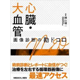
心臓・大血管 画像診断の勘ドコロNEO

出版社: メジカルビュー社
著者:
発行日: 2021-03-30
分野: 臨床医学:一般  >  画像/超音波
ISBN: 9784758316156
シリーズ: 画像診断の勘ドコロ
電子書籍版: 2021-03-30 (第1版第1刷)
書籍・雑誌
≪全国送料無料でお届け≫
取寄せ目安:約3営業日

6,820 円(税込)

電子書籍
章別単位での購入はできません
ブラウザ、アプリ閲覧

6,820 円(税込)

商品紹介

「勘ドコロNEO」シリーズ第5弾は,心臓・大血管の画像診断の要点をコンパクトにまとめた一冊。「どこから学び始めたら良いか,基本がわからない」「循環器疾患や末梢血管の診断レポートを知りたい」といった声に応える。エキスパートによるとっておきの画像をとともに,画像所見や鑑別点を端的に解説。目玉となる「ここが勘ドコロ」はもちろん,押さえておきたい文献やガイドラインを取り上げた「必読」,教科書にない経験知が凝縮された「ここだけの話」など欄外も充実。経験者にも役立つ入門書。

目次

  • 第1章 基礎編
    I 心臓CTのminimum requirements
     01.心臓CTの最新技術学
     02.心臓CTに親しむ

    II 心臓MRIのminimum requirements
     01.心臓MRIの最新撮像法
     02.心臓MRIに親しむ 

    III 心臓核医学のminimum requirements
     01.心臓核医学の最新撮像法
     02.心臓核医学に親しむ

    第2章 疾患編
    I 虚血性心疾患
     01.心筋虚血
     02.心筋梗塞

    II そのほかの冠動脈疾患
     01.冠動脈奇形
     02.冠動脈瘤
     03.心筋ブリッジ

    III 心筋症
     01.肥大型心筋症Ⅰ(HCM)
     02.肥大型心筋症Ⅱ(HOCM,心尖部肥大型心筋症,心中部肥大型心筋症)
     03.拡張型心筋症(DCM)
     04.左室緻密化障害(LVNC)
     05.不整脈原性右室心筋症(ARVC)
     06.心サルコイドーシス 
     07.ヘモクロマトーシス,ヘモジデローシス
     08.心アミロイドーシス
     09.心ファブリー病 
     10.膠原病関連の心病変
     11.たこつぼ心筋症
     12.高血圧性心筋症 

    IV 炎症性疾患
     01.急性心筋炎
     02.好酸球性心筋炎
     03.感染性心内膜炎(IE)
     04.収縮性心膜炎 

    V 腫瘍・腫瘤性病変
     01.粘液腫
     02.乳頭状弾性線維腫
     03.脂肪腫
     04.線維腫,横紋筋腫
     05.血管腫
     06.心腔内血栓
     07.血管肉腫
     08.悪性リンパ腫
     09.転移性心臓腫瘍
     10.心膜囊胞,心膜憩室

    VI 弁膜疾患
     01.大動脈弁狭窄症,大動脈二尖弁,TAVI/TAVR
     02.大動脈弁閉鎖不全症(AR)
     03.僧帽弁閉鎖不全症(MR)

    VII 先天性心疾患
     01.心房中隔欠損症
     02.卵円孔開存
     03.心室中隔欠損症
     04.Fallot四徴症
     05.Fontan循環
     06.三心房心(cor triatriatum sinistrum)
     07.分界稜(prominent crista terminalis)
     08.調節帯(moderator band)

    VIII 大血管
     01.大動脈瘤 
     02.バルサルバ洞動脈瘤(破裂)
     03.胸腹部大動脈瘤とAdamkiewicz動脈
     04.炎症性大動脈瘤
     05.感染性動脈瘤
     06.遺伝性結合組織疾患に伴う大動脈瘤,大動脈解離(Marfan症候群など)
     07.大動脈瘤切迫破裂
     08.大動脈瘤破裂
     09.大動脈瘤に対するステントグラフト留置術と術後評価(エンドリーク)
     10.大動脈解離 
     11.Penetrating atherosclerotic ulcer(PAU)
     12.大動脈弓の先天異常
     13.大動脈縮窄症
     14.大静脈の先天異常
     15.動脈管開存
     16.胸郭出口症候群
     17.高安動脈炎
     18.巨細胞性動脈炎
     19.結節性多発動脈炎
     20.腎血管性高血圧
     21.線維筋性異形成
     22.内臓動脈瘤
     23.正中弓状靱帯圧迫症候群(MALS)
     24.分節性動脈中膜融解症(SAM)
     25.腸間膜動脈閉塞症,解離
     26.非閉塞性腸間膜虚血(NOMI)

    IX 肺循環
     01.静脈血栓塞栓症(肺血栓塞栓症,深部静脈血栓症)
     02.肺動静脈奇形
     03.肺高血圧症
     04.肺静脈閉塞性疾患(PVOD)
     05.総肺静脈還流異常
     06.部分肺静脈還流異常(scimitar症候群を含む)

    X 末梢血管
     01.閉塞性動脈硬化症
     02.バージャー病
     03.下肢静脈瘤
     04.膝窩動脈捕捉症候群
     05.遺残坐骨動脈

この書籍の参考文献

参考文献のリンクは、リンク先の都合等により正しく表示されない場合がありますので、あらかじめご了承下さい。

本参考文献は電子書籍掲載内容を元にしております。

第1章 基礎編

P.2 掲載の参考文献
1. Schroeder S, et al:Cardiac computed tomography:indications, applications, limitations, and training requirements:Report of a Writing Group deployed by the Working Group Nuclear Cardiology and Cardiac CT of the European Society of Cardiology and the European Council of Nuclear Cardiology. Eur Heart J, 29:531-556, 2008.
2. 日本循環器学会:循環器病の診断と治療に関するガイドライン(2007-2008年度合同研究班報告):冠動脈病変の非侵襲的診断法に関するガイドライン. Circ J, 2009;73, Suppl III, 2009. http://www.j-circ.or.jp/guideline/pdf/JCS2010_yamashina_h.pdf
P.14 掲載の参考文献
3. Leipsic J, et al:CT angiography(CTA) and diagnostic performance of noninvasive fractional flow reserve:results from the Determination of Fractional Flow Reserve by Anatomic CTA (DeFACTO) study. AJR Am J Roentgenol, 202:989-994. PMID:24758651, 2014.
P.15 掲載の参考文献
4. 山口隆義:ファントムを用いたTDCの適正化について. MDCT至適造影法を語る会~64列心臓MDCTの造影法, 2008.
P.19 掲載の参考文献
Cardiac Imaging 2008. 臨床画像11月増刊号 24, 2008.
Cardiac Imaging 2015. 臨床画像4月増刊号 31, 2015.
日本循環器学会ほか:慢性冠動脈疾患診断ガイドライン(2018年改訂版). 52-62, 2019. http://www.j-circ.or.jp/guideline/pdf/JCS2018_yamagishi_tamaki.pdf
P.20 掲載の参考文献
1. Austen WG, et al:A reporting system on patients evaluated for coronary artery disease. Report of the AdHoc Committee for Grading of Coronary Artery Disease, Council on Cardiovascular Surgery, American Heart Association. Circulation, 51:5-40, 1975.
2. Raff GL, et al:SCCT guidelines for the interpretation and reporting of coronary computed tomographic angiography. J Cardiovasc Comput Tomogr, 3:122-136, 2009.
P.21 掲載の参考文献
3. Kyriakidis MK, et al:Sinus node coronary arteries studied with angiography. Am J Cardiol, 51:749-750, 1983.
P.23 掲載の参考文献
4. Saladin KS:The circulatory system;the heart. In:Anatomy and Physiology. The unity of form and function(5th ed). The McGraw-Hill Companies, New York, p742-744, 2010.
5. Fernandes JM, et al:Doppler-derived myocardial performance index in patients with impaired left ventricular relaxation and preserved systolic function. Echocardiography, 26:907-915, 2009.
6. Husmann L, et al:Coronary artery motion and cardiac phases:Dependency on heart rate-Implications for CT image reconstruction. Radiology, 245:567-557, 2007.
7. Okada M, et al:Systolic reconstruction in patients with low heart rate using coronary dual-source CT angiography. Eur J Radiol, 80:336-341, 2011.
P.24 掲載の参考文献
8. Abbara S, et al:SCCT guidelines for performance of coronary computed tomographic angiography:A report of the Society of Cardiovascular Computed Tomography Guidelines Committee. J Cardiovasc Comput Tomogr, 3:190-204, 2009.
P.25 掲載の参考文献
9. Budoff MJ, et al:Diagnostic performance of 64-multidetector row coronary computed tomographic angiography for evaluation of coronary artery stenosis in individuals without known coronary artery disease:results from the prospective multicenter ACCURACY(Assessment by Coronary Computed Tomographic Angiography of Individuals Undergoing Invasive Coronary Angiography)trial. J Am Coll Cardiol, 52:1724-1732, 2008.
10. Raff GL, et al:Diagnostic accuracy of noninvasive coronary angiography using 64-slice spiral computed tomography. J Am Coll Cardiol, 46:552-557, 2005.
11. Miller JM, et al:Diagnostic performance of coronary angiography by 64-row CT. N Engl J Med, 359:2324-2336, 2008.
12. Achenbach S, et al:Detection of calcified and noncalcified coronary atherosclerotic plaque by contrast-enhanced, submillimeter multidetector spiral computed tomography:a segment-based comparison with intravascular ultrasound. Circulation, 109:14-17, 2004.
P.26 掲載の参考文献
13. Cury RC, et al:Coronary artery disease-reporting and data system(CAD-RADS):An expert consensus document of SCCT, ACR and NASCI:endorsed by the ACC. JACC Cardiovasc Imaging, 9:1099-1113, 2016.
14. Xie JX, et al:The Coronary artery disease-reporting and data system(CAD-RADS):Prognostic and clinical implications associated with standardized coronary computed tomography angiography reporting. JACC Cardiovasc Imaging, 11:78-89, 2018.
P.27 掲載の参考文献
16. Schroeder S, et al:Noninvasive detection and evaluation of atherosclerotic coronary plaques with multislice computed tomography. J Am Coll Cardiol, 37:1430-1435, 2001.
17. Schroeder S, et al:Reliability of differentiating human coronary plaque morphology using contrast-enhanced multislice spiral computed tomography. A comparison with histology. J Comput Assist Tomogr, 28:449-454, 2004.
18. Leber AW, et al:Accuracy of multidetector spiral computed tomography in identifying and differentiating the composition of coronary atherosclerotic plaques. J Am Coll Cardiol, 43:1241-1247, 2004.
19. Viles-Gonzalez JF, et al:In vivo 16-slice, multidetector-row computed tomography for the assessment of experimental atherosclerosis. Comparison with magnetic resonance imaging and histopathology. Circulation, 110:1467-1472, 2004.
20. Sun J, et al:Identification and quantification of coronary atheriosclerotic plaques:Acomparison of 64-MDCT and intravascular ultrasound. AJR, 190:748-754, 2008.
P.29 掲載の参考文献
21. Greenland P, et al:ACCF/AHA 2007 clinical expert consensus document on coronary artery calcium scoring by computed tomography in global cardiovascular risk assessment and in evaluation of patients with chest pain. Circulation, 115:402-426, 2007.
22. Halliburton SS, et al:SCCT guidelines on radiation dose and dose-optimization strategies in cardiovascular CT. J Cardiovasc Comput Tomogr, 5:198-224, 2011.
P.30 掲載の参考文献
1. Kramer CM, et al:Standardized cardiovascular magnetic resonance(CMR)protocols 2020 update. J cardiovasc Magn Reson, 22:17, 2020.
2. Schulz-Menger J, et al:Standardized image interpretation and post processing in cardiovascular magnetic resonance:Society for Cardiovascular Magnetic Resonance(SCMR)Board of Trustees Task Force on Standardized Post Processing. J cardiovasc Magn Reson, 15:35, 2013.
3. Hundley WG, et al:Society for Cardiovascular Magnetic Resonance guidelines for reporting cardiovascular magnetic resonance examinations. J cardiovasc Magn Reson, 11:5, 2009.
P.33 掲載の参考文献
栗林幸夫ほか:心臓血管疾患のMDCTとMRI, 医学書院, 東京, 2005.
P.36 掲載の参考文献
4. Giri, S, et al:T2 quantification for improved detection of myocardial edema. J Cardiovasc Magn Reson, 11:56, 2009.
5. Sprinkart AM, et al:Gradient Spin Echo(GraSE)imaging for fast myocardial T2 mapping. J Cardiovasc Magn Reson, 17:12, 2015.
P.39 掲載の参考文献
6. Haaf P, et al:Cardiac T1 mapping and extracellular volume(ECV)in clinical practice:a comprehensive review. J Cardiovasc Magn Reson, 18:89-101, 2016.
P.41 掲載の参考文献
7. M. Ishida, et al:Impact of an abdominal belt on breathing patterns and scan efficiency in whole-heart coronary magnetic resonance angiography:comparison between the UK and Japan. J Cardiovasc Magn Reson, 13:71, 2011.
P.43 掲載の参考文献
1. Auster WG, et al:A Reporting System on Patients Evaluated for Coronary Artery Disease. Report of the Ad Hoc Committee for Grading of Coronary Artery Disease, Council on Cardiovascular Surgery, American Heart Association. Circulation, 51(4 suppl):5-40, 1975.
P.44 掲載の参考文献
2. Cerqueira MD, et al:Standardized Myocardial Segmentation and Nomenclature for Tomographic Imaging of the Heart. A Statement for healthcare Professionals From the Cardiac Imaging Commitee of the Council on Clinical Cardiology of the American Heart Association. Circulation, 105:539-542, 2002.
3. Greenwood JP, et al:Cardiovascular magnetic resonance and single-photon emission computed tomography for diagnosis of coronary heart disease(CE-MARC):a prospective trial. Lancet, 379:453-460, 2012.
P.45 掲載の参考文献
4. Kim RJ, et al:Relationship of MRI delayed contrast enhancement to irreversible injury, infarct age, and contractile function. Circulation, 100:1992-2002, 1999.
P.48 掲載の参考文献
5. Messroghli DR, et al:Clinical recommendations for cardiovascular magnetic resonance mapping of T1, T2, T2*and extracellular volume:A consensus statement by the Society for Cardiovascular Magnetic Resonance(SCMR)endorsed by the European Association for Cardiovascular Imaging(EACVI). J Cardiovasc Magn Reson, 19:75, 2017.
栗林幸夫ほか:心臓血管疾患のMDCTとMRI, 医学書院, 東京, 2005.
クラウス・D. クラウセンほか:わかる! 心臓画像診断の要点(似鳥俊明 監訳), メディカル・サイエンス・インターナショナル, 東京, 2009.
P.49 掲載の参考文献
1. Anger H O:SCINTILLATION CAMERA WITH MULTICHANNEL COLLIMATORS. J Nucl Med, 5:515-531, 1964.
2. Gambhir SS, et al:A novel high-sensitivity rapid-acquisition single-photon cardiac imaging camera. J Nucl Med, 50:635-643, 2009.
P.53 掲載の参考文献
3. Fiechter M, et al:Diagnostic value of 13N-ammonia myocardial perfusion PET:added value of myocardial flow reserve. J Nucl Med, 53:1230-12344, 2012.
4. Nishiyama Y, et al:Combined supine and prone myocardial perfusion single-photon emission computed tomography with a cadmium zinc telluride camera for detection of coronary artery disease. Circ J, 78:1169-1175, 2014.
P.60 掲載の参考文献
1. Hachamovitch R, et al:Comparison of the short-term survival benefit associated with revascularization compared with medical therapy in patients with no prior coronary artery disease undergoing stress myocardial perfusion single photon emission computed tomography. Circulation, 107:2900-2907, 2003.
2. Nishimura T, et al:Prognostic study of risk stratification among Japanese patients with ischemic heart disease using gated myocardial perfusion SPECT:J-ACCESS study. Eur J Nucl Med Mol Imaging, 35:319-328, 2008.
P.61 掲載の参考文献
久保敦司ほか:IV. 循環器. 核医学ノート, 第6版, 123-213, 金原出版, 東京, 2019.
橋本 順:A心臓 核医学の基礎. 心筋SPECT, 西村恒彦ほか編著, 9-38, 金原出版, 東京, 2012.

第2章 疾患編

P.64 掲載の参考文献
1. Knuuti J, et al:2019 ESC Guidelines for the diagnosis and management of chronic coronary syndromes:The Task Force for the diagnosis and management of chronic coronary syndromes of the European Society of Cardiology(ESC). European Heart Journal, 2019.
2. Hachamovitch R, et al:Comparison of the short-term survival benefit associated with revascularization compared with medical therapy in patients with no prior coronary artery disease undergoing stress myocardial perfusion single photon emission computed tomography. Circulation, 107:2900-2907, 2003.
3. Manisty C, et al:Splenic Switchoff:A Tool to Assess Stress Adequacy in Adenosine Perfusion Cardiac MR Imaging. Radiology, 276:732-740, 2015.
P.66 掲載の参考文献
4. Karamitsos TD, et al:Feasibility and safety of high-dose adenosine perfusion cardiovascular magnetic resonance. J Cardiovasc Magn Reson, 12:66, 2010.
5. Kramer CM, et al:Society for Cardiovascular Magnetic Resonance Board of Trustees Task Force on Standardized P. Standardized cardiovascular magnetic resonance(CMR)protocols 2013 update. J Cardiovasc Magn Reson, 15:91, 2013.
日本循環器学会ほか:慢性冠動脈疾患診断ガイドライン(2018年改訂版).
P.67 掲載の参考文献
1. 高久史麿ほか監訳:ワシントンマニュアル第13版, メディカル・サイエンス・インターナショナル, 東京, 2015.
西村恒彦編:BRAND NEW心臓核医学-機能画像が病態を捉える, 金原出版, 東京, 2012.
SCMR Japan Working Groupホームページ. http://scmr.jp/scmrjc/
P.69 掲載の参考文献
2. Kim RJ, et al:The use of contrast-enhanced magnetic resonance imaging to identify reversible myocardial dysfunction. N Engl J Med, 343:1445-1453, 2000.
P.71 掲載の参考文献
1. Maron BJ, et al:Sudden death in young competitive athletes:clinical, demographic, and pathological profiles. JAMA, 276:199-204, 1996.
2. Taylor AJ, et al:Sudden cardiac death associated with isolated congenital coronary artery anomalies. J Am Coll Cardiol, 20:640-647, 1992.
3. Angelini P, et al:Coronary anomalies:incidence, pathophysiology, and clinical relevance. Circulation, 105:2449-2454, 2002.
P.74 掲載の参考文献
1. Manginas A, et al:Coronary artery ectasias:imaging, functional assessment and clinical implications. Eur Heart J, 27:1026-1031, 2006.
2. 日本循環器学会ほか:循環器病の診断と治療に関するガイドライン(2012年度合同研究班報告)川崎病心臓血管後遺症の診断と治療に関するガイドライン(2013年改訂版).
P.76 掲載の参考文献
1. Ishikawa Y, et al:Histopathologic profiles of coronary atherosclerosis by myocardial bridge underlying myocardial infarction. Atherosclerosis, 226:118-123, 2013.
2. Zeina AR, et al:Myocardial bridge:evaluationon MDCT. AJR, 88:1069-1073, 2007.
3. Ishii T, et al:The effects of a myocardial bridge on coronary artherosclerosis and ischaemia. J Pathol, 185:4-9, 1998.
P.78 掲載の参考文献
1. Amano Y, et al:Cardiac MR imaging of hypertrophic cardiomyopathy:techniques, findings, and clinical relevance. Magn Reson Med Sci, 17:120-131, 2018.
2. Maron MS:Clinical utility of cardiovascular magnetic resonance in hypertrophic cardiomyopathy. J Cardiovasc Magn Reson, 14:13, 2012.
難病情報センター. http://www.nanbyou.or.jp/entry/177
P.80 掲載の参考文献
1. Amano Y, et al:Cardiac MR imaging of hypertrophic cardiomyopathy:techniques, findings, and clinical relevance. Magn Reson Med Sci, 17:120-131, 2018.
2. Amano Y, et al:Delayed-enhancement MRI of apical hypertrophic cardiomyopathy:assessment of the intramural distribution and comparison with clinical symptoms, ventricular arrhythmias, and cine MRI. ActaRadiol, 52:613-618, 2011.
難病情報センターhttp://www.nanbyou.or.jp/entry/177
P.82 掲載の参考文献
1. McCrohon JA, et al:Differentiation of Heart Failure Related to Dilated Cardiomyopathy and Coronary Artery Disease Using Gadolinium-Enhanced Cardiovascular Magnetic Resonance. Circulation, 108:54-59, 2003.
2. Gulati A, et al:Association of Fibrosis With Mortality and Sudden Cardiac Death in Patients With Nonischemic Dilated Cardiomyopathy. JAMA, 309:896-908, 2013.
3. Yanagisawa F, et al:Non-con-trast-enhanced T1 Mapping of Dilated Cardiomyopathy:Comparison between Native T1 Values and Late Gadolinium Enhancement. Magn Reson Med Sci, 18:12-18, 2019.
難病情報センターhttp://www.nanbyou.or.jp/entry/177
P.84 掲載の参考文献
1. Feldt RH, et al:Anomalous ventricular myocardial patterns in a child with complex congenital heart disease. Am J Cardiol, 23:732-734, 1969.
2. Chin TK, et al:Isolated Noncompaction of Left Ventricular Myocardium A study of Eight Cases. Circulation, 82:507-513, 1990.
3. Wang C, et al:Long-Term Prognosis of Patients With Left Ventricular Noncompaction-Comparison Between Infantile and Juvenile Types. Cic J, 81:694-700, 2017.
4. Andreini D, et al:Long-Term Prognostic Value of Cardiac Magnetic Resonance in Left Ventricule Noncompaction. A Prospective Multicenter Study. J Am Coll Cardiol, 68:2166-2181, 2016.
P.85 掲載の参考文献
5. Petersen J, et al:Left Ventricular Non-Compaction Insights From Cardiovascular Magnetic Resonance Imaging. Am Coll Cardiol, 46:101-105, 2005.
6. Kawel N, et al:Trabeculated(Noncompacted)and Compact Myocardium in Adults:The Multi-Ethnic Study of Atherosclerosis. Circ Cardiovasc Imaging, 5:357-66, 2012.
7. Stacey BR, et al:Comparison of Systolic and Diastolic Criteria for Isolated LV Noncompaction in CMR. JACC Cardiovasc Imaging, 6:931-940, 2013.
8. Jacquier A, et al:Measurement of Trabeculated Left Ventricular Mass Using Cardiac Magnetic Resonance Imaging in the Diagnosis of Left Ventricular Non-Compaction. Eur Heart J, 31:1098-104, 2010.
9. Weir-McCall JR, et al:Left Ventricular Noncompaction:Anatomical Phenotype or Distinct Cardiomyopathy? J Am Coll Cardiol, 68:2157-2165, 2016.
P.86 掲載の参考文献
1. Marcus FI, et al:Diagnosis of arrhythmogenic right ventricular cardiomyopathy/dysplasia:Proposed modification of the task force criteria. Circulation, 1 21:1533-1541, 2010.
P.87 掲載の参考文献
2. Femia G, et al:Long term CMR follow up of patients with right ventricular abnormality and clinically suspected arrhythmogenic right ventricular cardiomyopathy(ARVC). J Cardiovasc Magn Reson, 21:76, 2019.
3. Melak LA, et al:Cardiovascular magnetic resonance with parametric mapping in long-term ultra-marathon runners. Eur J Radiol, 117:89-84, 2019.
4. Matsuo Y, et al:CT Features of Myocardial Fat and Correlation With Clinical Background in Patients Without Cardiac Disease. Jpn J Radiol, 31:444-454, 2013.
Kimura F, et al:Myocardial Fat at Cardiac Imaging:How Can We Differentiate Pathologic from Physiologic Fatty Infiltration? Radiographics, 30:1587-1602, 2010.
P.88 掲載の参考文献
1. 森本泰介ほか:2004年サルコイドーシス疫学調査. 日サ会誌, 27:103-108, 2007.
2. Iwai K, et al:Pathological studies on sarcoidosis autopsy. I. Epidemiological features of 320 cases in Japan. Acta Pathol Jpn, 43:372-376, 1993.
3. Puntmann VO, et al:T1 and T2 mapping in recognition of early cardiac involvement in systemic sarcoidosis. Radiology, 285:63-72, 2017.
P.90 掲載の参考文献
4. 日本循環器学会:2016年版心臓サルコイドーシスの診療ガイドライン. www.j-circ.or.jp/guideline/pdf/JCS2016_terasaki_h.pdf
5. Ohira H, et al:Inter-and Intraobserver Agreement of 18F-FDG PET/CT Image Interpretation in Patients Referred for Assessment of Cardiac Sarcoidosis. J Nucl Med, 58:1324-1329, 2017.
6. Kumita S, et al:Recommendations for 18F-fluorodeoxyglucose positron emission tomography imaging for diagnosis of cardiac sarcoidosis-2018 update:Japanese Society of Nuclear Cardiology recommendations. J Nucl Cardiol, 26:1414-1433, 2019.
日本心臓核医学会:心臓サルコイドーシスに対する18FFDG PET検査の手引き2018年度改訂. 心サルコイドーシスのPETの診断のガイドライン心臓核医会誌, 21:22-27, 2019.
P.91 掲載の参考文献
1. Gujja P, et al:Iron Overload Cardiomyopathy Better Understanding of an Increasing Disorder. J Am Coll Cardiol, 56:1001-1012, 2010.
P.92 掲載の参考文献
2. Messroghli DR, et al:Clinical recommendations for cardiovascular magnetic resonance mapping of T1, T2, T2*and extracellular volume:A consensus statement by the Society for Cardiovascular Magnetic Resonance(SCMR)endorsed by the European Association for Cardiovascular Imaging(EACVI). J Cardiovasc Magn Reson, 19:75, 2017.
3. Hanneman K, et al:Quantification of Myocardial Extracellular Volume Fraction with Cardiac MR Imaging in Thalassemia Major. Radiology, 279:720-730, 2016.
4. Kirk P, et al:Low prevalence of fibrosis in thalassemia major assessed by late gadolinium enhancement cardiovascular magnetic resonance. J Cardiovasc Magn Reson, 13:8, 2011.
P.93 掲載の参考文献
日本循環器学会:心アミロイドーシス診療ガイドライン, 2020年版
P.94 掲載の参考文献
1. Dorbala S, et al:ASNC/AHA/ASE/EANM/HFSA/ISA/SCMR/SNMMI expert consensus recommendations for multimodality imaging in cardiac amyloidosis:Part 1 of 2-evidence base and standardized methods of imaging. J Nucl Cardiol, 26:2065-2123, 2019.
2. Dorbala S, et al:ASNC/AHA/ASE/EANM/HFSA/ISA/SCMR/SNMMI expert consensus recommendations for multimodality imaging in cardiac amyloidosis:Part 2 of 2-Diagnostic criteria and appropriate utilization. J Nucl Cardiol, 26:2065-2123, 2019.
3. Syed IS, et al:Role of cardiac magnetic resonance imaging in the detection of cardiac amyloidosis. JACC Cardiovasc imaging, 3:155-164, 2010.
4. Fontana M, et al:Native T1 mapping in transthyretin amyloidosis. JACC Cardiovasc Imaging, 7:157-165, 2014.
5. Castano A, et al. Multicenter Study of Planar Technetium 99m Pyrophosphate Cardiac Imaging:Predicting Survival for Patients With ATTR Cardiac Amyloidosis. JAMA cardiology, 1:880-889, 2016.
6. Gillmore JD, et al:Nonbiopsy Diagnosis of Cardiac Transthyretin Amyloidosis. Circulation, 133:2404-2412, 2016.
P.96 掲載の参考文献
1. Inoue T, et al:Newborn Screening for Fabry Disease in Japan:Prevalence and Genotypes of Fabry Disease in a Pilot Study. J Hum Genet, 58:548-552, 2013.
2. Nakao S, et al:An Atypical Variant of Fabry's Disease in Men With Left Ventricular Hypertrophy. N Engl J Med, 333:288-293, 1995.
Messroghli D, et al:Clinical Recommendations for Cardiovascular Magnetic Resonance Mapping of T1, T2, T2* and Extracellular Volume:A Consensus Statement by the Society for Cardiovascular Magnetic Resonance (SCMR) Endorsed by the European Association for Cardiovascular Imaging(EACVI). J CMR, 19:75, 2017.
P.99 掲載の参考文献
Mavrogeni SI, et al:Cardiovascular magnetic resonance in rheumatology:Current status and recommendations for use. Int J Cardiol, 217:135-148, 2016.
P.100 掲載の参考文献
1. Greulichs, et al:T1 and T2 Mapping for Evaluation of Myocardial Involvement in Patients With ANCA-associated Vasculitides. Cardiovasc Magn Reson, 19:6, 2017.
P.102 掲載の参考文献
1. Kawai S,et al:Takotsubo Cardiomyopathy Group:Guidelines for diagnosis of takotsubo(ampulla)cardiomyopathy. Circ J, 71:990-992, 2007.
2. Izumo M, et al:Mechanisms of acute mitral regurgitation in patients with takotsubo cardiomyopathy:an echocardiographic study. Circ Cardiovasc Imaging, 4:392-398, 2011.
P.103 掲載の参考文献
3. Owa M, et al:Emotional stress-induced‘ampulla cardiomyopathy':discrepancy between the metabolic and sympathetic innervation imaging performed during the recovery course. Jpn Circ J, 65:349-352, 2001.
4. Matsuo S, et al:Diagnostic utility of 123I-BMIPP imaging in patients with Takotsubo cardiomyopathy. J Cardiol, 64:49-56, 2014.
5. Ghadri JR, et al:International Expert Consensus Document on Takotsubo Syndrome(Part II):Diagnostic Workup, Outcome, and Management. EUR Heart J, 39:2047-2062, 2018.
P.105 掲載の参考文献
1. 日本循環器学会:拡張型心筋症ならびに関連する二次性心筋症の診療に関するガイドライン, 2011.
2. Bohl S, et al:Delayed enhancement cardiac magnetic resonance imaging reveals typical patterns of myocardial injury in patients with various forms of non-ischemic heart disease. Int J Cardiovasc Imaging, 24:597-607,2008.
P.106 掲載の参考文献
3. Hinojar R, et al:T1 Mapping in Discrimination of Hypertrophic Phenotypes:Hypertensive Heart Disease and Hypertrophic Cardiomyopathy:Findings From the International T1 Multicenter Cardiovascular Magnetic Resonance Study. Circ Cardiovasc Imaging, 8:e003285, 2015.
4. Martinez-Naharro A, et al:Magnetic Resonance in Transthyretin Cardiac Amyloidosis. J Am Coll Cardiol, 70:466-477, 2017.
5. Sado DM, et al:Identification and assessment of Anderson-Fabry disease by cardiovascular magnetic resonance noncontrast myocardial T1 mapping. Circ Cardiovasc Imaging, 6:392-398, 2013.
P.108 掲載の参考文献
1. Friedrich MG, et al:Cardiovascular magnetic resonance in myocarditis:A JACC White Paper. JACC, 53:1475, 2009.
2. Ferreira VM, et al:Cardiovascular Magnetic Resonance in Nonischemic Myocardial Inflammation:Expert Recommendations. JACC, 72:3158, 2018.
日本循環器学会ほか:急性および慢性心筋炎の診断・治療に関するガイドライン(2009年改訂版).www.j-circ.or.jp/guideline/pdf/JCS2009_izumi_h.pdf
P.110 掲載の参考文献
1. Morimoto S, et al:Changes in the peripheral eosinophil count in patients with acute eosinophilic myocarditis. Heart Vessels, 18:193-196, 2003.
2. Mori N, et al:Clinical pictures of 35 cases with eosinophilic myocarditis. Circ J, 68(Suppl I):244(abstr), 2004.
3. Yanagisawa T, et al:Clinical Significance of Corticosteroid Therapy for Eosinophilic Myocarditis.Int Heart J, 52:110-113, 2011.
P.113 掲載の参考文献
1. 日本循環器学会ほか:感染性心内膜炎の予防と治療に関するガイドライン(2017年改訂版), 2019
P.115 掲載の参考文献
1. Cosyns B, et al:European Association of Cardiovascular Imaging(EACVI)position paper:multimodality imaging in pericardial disease. Eur Heart J Cardiovasc Imaging, 16:12-31, 2015.
2. Alajaji W, et al:Noninvasive multimodality imaging for the diagnosis of constrictive pericarditis. Circ Cardiovasc Imaging, 11:11e007878, 2018.
Adler Y, et al:2015 ESC Guidelines for the diagnosis and management of pericardial diseases:The Task Force for the Diagnosis and Management of Pericardial Diseases of the European Society of Cardiology(ESC)Endorsed by:The European Association for Cardio-Thoracic Surgery(EACTS). Eur Heart J, 36:2921-2964, 2015.
P.117 掲載の参考文献
1. Shetty Roy AN, et al:Familial recurrent atrial myxoma:Carney's complex. Clin Cardiol, 34:83-86, 2011.
2. Colin GC, et al:Cardiac myxoma:a contemporary multimodality imaging review. Int J Cardiovasc Imaging, 34:1789-1808, 2018.
P.118 掲載の参考文献
3. Hong YJ, et al:Dual-energy cardiac computed tomography for differentiating cardiac myxoma from thrombus. Int J Cardiovasc Imaging, 30 Suppl 2:121-128, 2014.
P.119 掲載の参考文献
1. Sun JP, et al:Clinical and echocardiographic characteristics of papillary fibroelastomas:a retrospective and prospective study in 162 patients. Circulation, 103:2687-2693, 2001.
2. Gowda RM, et al:Cardiac papillary fibroelastoma:a comprehensive analysis of 725 cases. Am Heart J, 146:404-410, 2003.
3. Motwani M, et al:MR imaging of cardiac tumors and masses:a review of methods and clinical applications. Radiology, 268:26-43, 2013.
4. Sparrow PJ, et al:MR imaging of cardiac tumors. Radiographics, 25:1255-1276, 2005.
P.120 掲載の参考文献
5. Tamin SS, et al:Prognostic and Bioepidemiologic Implications of Papillary Fibroelastomas. J Am Coll Cardiol, 65, 2420-2429, 2015.
P.121 掲載の参考文献
1. Grebenc ML, et al:Primary cardiac and pericardial neoplasms:radiologic-pathologic correlation. Radiographics, 20:1073-1103, 2000.
2. Sparrow PJ, et al:MR imaging of cardiac tumors. Radiographics, 25:1255-1276, 2005.
3. Kassop D, et al:Cardiac masses on cardiac CT:a review. Curr Cardiovasc Imaging Rep, 7:9281, 2014.
P.122 掲載の参考文献
4. Cannavale G, et al:Fatty images of the heart:spectrum of normal and pathological findings by computed tomography and cardiac magnetic resonance imaging. Biomed Res Int 5610347, 2018.
P.123 掲載の参考文献
5. Shaaya EA, et al:Cardiac fat-containing lesions are common in tuberous sclerosis complex. Am J Med Genet A, 16:1662-1665, 2013.
P.124 掲載の参考文献
1. Tao TY, et al:Pediatric cardiac tumors:clinical and imaging features:Radiographics, 34:1031-1046, 2014.
2. Nwachukwu H, et al:Cardiac fibroma in adults. Cardiovasc Pathol, 20:e146-e152, 2011.
3. Grebenc ML, et al:Primary cardiac and pericardial neoplasms:radiologic-pathologic correlation. Radiographics, 20:1073-1103;quiz 1110-1112, 2000.
P.126 掲載の参考文献
4. Sparrow PJ, et al:MR imaging of cardiac tumors:Radiographics, 25:1255-1276, 2005.
P.127 掲載の参考文献
1. Weidong Li, et al:Cardiac Hemangioma:A Comprehensive Analysis of 200 Cases. Ann Thorac Surg, 99:2246-2252, 2015.
2. Kojima S, et al:Cardiac hemangioma:2 report of two cases and review of the literature. Heart Vessels, 18:153-156, 2003.
3. Grenbenc ML, et al:Primary cardiac and pericardial neoplasms:radiographic correlation. Radiographics, 20:1073-1103, 2000.
4. Sparrow PJ, et al:MR imaging of Cardiac Tumors. Radiographics, 25:1255-1276, 2005.
P.129 掲載の参考文献
1. McCarthy CP, et al:Left Ventricular Thrombus After Acute Myocardial Infarction:Screening, Prevention, and Treatment. JAMA Cardiol, 3:642-649, 2018.
2. Romero J, et al:Cardiac imaging for assessment of left atrial appendage stasis and thrombosis. Nat Rev Cardiol, 11:470-480, 2014.
3. Bulluck H, et al:Incidence and predictors of left ventricular thrombus by cardiovascular magnetic resonance in acute ST-segment elevation myocardial infarction treated by primary percutaneous coronary intervention:a meta-analysis. J Cardiovasc Magn Reason, 20:72, 2018.
吉原 修ほか:心臓CTを用いた心原性脳塞栓症の原因心疾患の評価. 心臓, 46:1399-1407, 2014.
P.132 掲載の参考文献
1. Araoz PA, et al:CT and MR imaging of primary cardiac malignancies. Radiographics, 19:1421-1434, 1999.
2. Sparrow PJ, et al:MR imaging of cardiac tumors. Radiographics, 25:1255-1276, 2005.
3. Deetjen AG, et al:Cardiac angiosarcoma diagnosed and characterized by cardiac magnetic resonance imaging. Cardiol Rev, 14:101-103, 2006.
4. Aoka Y, et al:Primary cardiac angiosarcoma treated with carbon-ion radiotherapy. Lancet Oncol, 5:636-638, 2004.
P.133 掲載の参考文献
5. Kim EE, et al:Malignant cardiac fibrous histiocytomas and angiosarcomas:MR features. J Comput Assist Tomogr, 13:627-632, 1989.
6. Yahata S, et al:Sunray appearance on enhanced magnetic resonance image of cardiac angiosarcoma with pericardial obliteration. AM Heart J, 127:468-471, 1994.
P.134 掲載の参考文献
7. Yu J, et al:Clinical and imaging manifestations of primary cardiac angiosarcoma. BMC Medical Imaging, 19:16, 2019.
P.135 掲載の参考文献
1. Randhawa K, et al:Magnetic resonance imaging of cardiac tumors:Part 2, malignant tumors and tumor-like conditions. Curr Probl Diagn Radiol, 40:169-179, 2011.
2. Miguel CE, et al:Primary cardiac lymphoma. International Journal of cardiology, 149:358-363, 2011.
3. Kikuchi Y, et al:Imaging characteristics of cardiac dominant diffuse large B-cell lymphoma demonstrated with MDCT and PET/CT. Eur J Nucl Med Mol Imaging, 40:1337-1344, 2013.
4. Page M, et al:Primary cardiac lymphoma:Diagnosis and the impact of chemotherapy on cardiac structure and function. Can J Cardiol, 32:931e1-e3, 2016.
P.136 掲載の参考文献
5. Dorsay TA, et al:Primary cardiac lymphoma:CT and MR findings. J Comput Assist Tomogr, 17:978-981, 1993.
6. Carter BW, et al:Multimodality imaging of cardiothoracic lymphoma. Eur Radiol, 83:1470-1482, 2014.
P.137 掲載の参考文献
7. Colin GC, et al:Value of CMR to differentiate cardiac angiosarcoma from cardiac lymphoma. JACC Cardiovasc Imaging, 8:744-746, 2015.
P.138 掲載の参考文献
1. Bussani R, et al:Cardiac metastases. J Clin Pathol, 60:27-34, 2007.
2. Chiles C, et al:Metastatic involvement of the heart and pericardium:CT and MR imaging. Radiographics, 21:439-449, 2001.
3. Puranik AD, et al:Asymptomatic myocardial metastasis from cancers of upper aero-digestive tract detected on FDG PET/CT:a series of 4 cases. Cancer Imaging, 14:16, 2014.
P.140 掲載の参考文献
1. 寺松 考ほか:縦隔腫瘍に関する全国集計-第1篇 縦隔腫瘍全国集計-. 日胸学会誌, 24:264-269, 1996.
2. 松下葉子ほか:心室憩室の2症例. 日胸疾会誌, 30:1842, 1992.
3. 森田久樹:心膜嚢胞. 別冊日本臨床新領域別症候群シリーズNo. 5 循環器症候群-その他の疾患を求めて, 435-437, 日本臨牀社, 東京, 1996.
4. King JF, et al:Rupture of pericardial cyst. Chest, 60:611-612, 1971.
5. Kruger SR, et al:Spontaneous resolution of pericardial cyst. Am Heart J, 109:1390-1391, 1985.
6. 横山健一ほか:心膜嚢胞とpericardial cystと右心横隔膜角の嚢胞性腫瘤. 画像診断(臨増), 32:82-83, 2012.
P.143 掲載の参考文献
1. Nishimura RA, et al:2014 AHA/ACC Guideline for the Management of Patients With Valvular Heart Disease:a report of the American College of Cardiology/American Heart Association Task Force on Practice Guidelines. Circulation, 129:e521-643, 2014.
2. Ross J, Jr., et al:Aortic stenosis. Circulation, 38(1 Suppl):61-67, 1968.
P.145 掲載の参考文献
3. Achenbach S, et al:SCCT expertconsensus document on computed tomography imaging before transcatheter aortic valve implantation(TAVI)/transcatheter aortic valve replacement(TAVR). J Cardiovasc Comput Tomogr, 6:366-380, 2012.
4. Blanke P, et al:Computed tomography imaging in the context of transcatheter aortic valve implantation(TAVI)/transcatheter aortic valve replacement(TAVR):An expert consensus document of the Society of Cardiovascular Computed Tomography. J Cardiovasc Comput Tomogr. 13:1-20, 2019.
P.146 掲載の参考文献
5. Gilard M, et al:Registry of transcatheter aortic-valve implantation in highrisk patients. N Engl J Med, 366:1705-1715, 2012.
6. Osnabrugge RL, et al:Aortic stenosis in the elderly:disease prevalence and number of candidates for transcatheter aortic valve replacement:a meta-analysis and modeling study. J Am Coll Cardiol, 62:1002-1012, 2013.
P.149 掲載の参考文献
1. 大北 裕ほか:弁膜疾患の非薬物治療に関するガイドライン(2012年改訂版), 日本循環器学会, 2012.
2. Bonow R, et al:Aortic regurgitation(in Chapter 57 Valvular Heart Disease). Braunwald's Heart disease:A textbook of cardiovascular medicine, 7th ed, Elsevier Saunders, Philadelphia, 1592-1601, 2005.
3. Feigenbaum H:Aortic valve disease. In:Feigenbaum H editor. Echocardiography, 6th ed, Lippincott Williams & Wilkins, Philadelphia, 271-305, 2005.
4. Detaint D, et al:Quantitative echocardiographic determinants of clinical outcome in asymptomatic patients with aortic regurgitation:a prospective study. JACC Cardiovasc Imaging, 1:1-11, 2008.
5. Koo HJ, et al:Functional classification of aortic regurgitation using cardiac computed tomography:comparison with surgical inspection. Int J Cardiovasc Imaging, 34:1295-1303, 2018.
P.150 掲載の参考文献
6. Lee JC, et al:Evaluation of aortic regurgitation with cardiac magnetic resonance imaging:a systematic review. Heart, 104:103-110, 2018.
P.152 掲載の参考文献
1. 大北 裕ほか:弁膜疾患の非薬物治療に関するガイドライン(2012年改訂版), 日本循環器学会, 2012.
2. Bonow RO, et al:Guidelines for the management of patients with valvular heart disease:executive summary. A report of the American College of Cardiology/American Heart Association Task Force on Practice Guidelines(Committee on Management of Patients with Valvular Heart Disease). Circulation, 98:1949-1984, 1998.
3. Weir-McCall, et al:Mitral Valve Imaging with CT:Relationship with Transcatheter Mitral Valve Interventions. Radiology, 288:638-655, 2018.
4. Liu B, et al:The evolving role of cardiac magnetic resonance in primary mitral regurgitation:ready for primetime? Eur Heart J cardiovasc Imaging, 20:123-130, 2019.
P.153 掲載の参考文献
5. Calkoen EE, et al:Characterization and quantification of dynamic eccentric regurgitation of the left atrioventricular valve after atrioventricular septal defect correction with 4D flow cardiovascular magnetic resonance and retrospective valve tracking. J Cardiovasc Magn Reson, 17:1-9, 2012.
P.154 掲載の参考文献
1. Dahnert W:Radiology Review Manual(8th ed), Wolters Kluwer, Lippincot Williams & Wilkins. Philadelphia, 2017.
2. 高橋長裕:図解先天性心疾患血行動態の理解と外科治療第2版, 医学書院, 東京, 2014.
P.155 掲載の参考文献
3. Simon N, et al:CT imaging feature of artrioventricular shunts;what the radiologist must know. Insights Imaging, 7:119-129, 2016.
P.158 掲載の参考文献
1. Young JK, et al:Patent Foremen Ovale:Diagnosis with Multidetector CT-Comparison with Transesopjageal Echocardiography. Radiology, 250:61-67, 2009.
2. Simon N, et al:CT imaging feature of artrioventricular shunts;what the radiologist must know. Insights Imaging, 7:119-129, 2016.
P.160 掲載の参考文献
1. 高橋長裕:図解先天性心疾患血行動態の理解と外科治療(第2版), 医学書院, 東京, 2014.
2. Dahnert W:Radiology Review Manual(8th ed), Wolters Kluwer, Lippincot Williams & Wilkins, Philadelphia, 2017.
P.161 掲載の参考文献
3. Simon N, et al:CT imaging feature of artrioventricular shunts;what the radiologist must know. Insights Imaging, 7:119-129, 2016.
P.164 掲載の参考文献
1. Gatzoulis MA, et al:Depolarization-repolarization inhomogeneity after repair of tetralogy of Fallot. Circulation, 95:401-404, 1997.
2. Oosterhof T, et al:Preoperative thresholds of pulmonary valve replacement in patients with corrected tetralogy of Fallot. Circulation, 116:545-551, 2007.
3. Therrien J, et al:Impact of pulmonary valve replacement on arrhythmia propensity late after repair of tetralogy of Fallot. Circulation, 103:2489-2494, 2001.
4. Niwa K, et al:Progressive aortic root dilatation in adults late after repair of tetralogy of Fallot. Circulation, 106:1374-1378, 2002.
5. Yamasaki Y, et al:Quantitative assessment of right ventricular function and pulmonary regurgitation in surgically repaired tetralogy of Fallot using 256-slice CT:comparison with 3-Tesla MRI. Eur Radiol, 24:3289-3299, 2014.
P.166 掲載の参考文献
6. Shiina Y, et al:The relationship between extracellular volume fraction in symptomatic adults with tetralogy of Fallot and adverse cardiac events. J Cardiol, 75:424-431, 2020.
P.168 掲載の参考文献
1. Diller GP, et al:Survival prospects and circumstances of death in contemporary adult congenital heart disease patients under follow-up at a large tertiary centre. Circulation, 132:2118-2125, 2015.
P.169 掲載の参考文献
2. Ishizaki U, et al:Global strain and dyssynchrony of the single ventricle predict adverse cardiac events after the Fontan procedure:analysis using feature-tracking cine magnetic resonance imaging. J Cardiol, 73:163-170, 2019.
P.170 掲載の参考文献
3. Ishizaki U, et al:Prediction of Fontan-associated liver disease using a novel cine magnetic resonance imaging"vortex flow map"in the right atrium. Circ J, 82:2143-2151, 2018.
4. Itkin M, et al:Protein-losing enteropathy in patients with congenital heart disease. J Am Coll Cardiol. 69:2929-2937, 2017.
P.171 掲載の参考文献
1. Van Son JA, et al:Cor triatriatum:diagnosis, operative approach, and late results. Mayo Clin Proc, 68:854-859, 1993.
2. Bartel T, et al:Dynamic three-dimensional echocardiography using parallel slicing:a promising diagnostic procedure in adults with congenital heart disease. Cardiology, 89:140-147, 1998.
P.173 掲載の参考文献
1. Broderick LS, et al:Anatomic pitfalls of the heart and pericardium. Radiographics, 25:Figure1, 441, 2005.
2. Mirowitz SA, et al:Fibromuscular elements of the right atrium:pseudomass at MR imaging. Radiology, 182:231-233, 1992.
3. Saremi F, et al:Cardiac conduction system:anatomic landmarks relevant to interventional electrophysiologic techniques demonstrated with 64-detector CT. Radiographics, 27:1539-1565, 2007.
4. Maurer AH, et al:How to differentiate benign versus malignant cardiac and paracardiac 18F FDG uptake at oncologic PET/CT. Radiographics, 31:1287-1305, 2011.
Malik SB, et al:Transthoracic echocardiography:pitfalls and limitations as delineated at cardiac CT and MR imaging. Radiographics, 37:383-406, 2017.
P.175 掲載の参考文献
1. Loukas M, et al:Anatomical observations of the moderator band. Clin Anat, 23:443-450, 2010.
2. Haddad F, et al:Right ventricular function in cardiovascular disease, part I:Anatomy, physiology, aging, and functional assessment of the right ventricle. Circulation, 117:1436-1448, 2008.
3. Uehara M, et al:Clinical significance of fat infiltration in the moderator band and right ventricular myocardium in multislice CT, and its association with abnormal conduction seen in electrocardiogram. Int J Cardiol, 168:352-256, 2013.
米澤政人ほか:右室二腔症. 画像診断別冊KEY BOOKシリーズ これだけは知っておきたい心臓・血管疾患の画像診断, 宇都宮大輔編著, 150-151, 秀潤社, 東京, 2016.
P.177 掲載の参考文献
1. 荻野 均ほか:大動脈瘤・大動脈解離診療ガイドライン(2020年改訂版) https://www.j-circ.or.jp/cms/wp-content/uploads/2020/07/JCS2020_Ogino.pdf
2. Chaikof EL, et al:The Society for Vascular Surgery practice guidelines on the care of patients with an abdominal aortic aneurysm. J Vasc Surg, 67:2-77.e2, 2018.
P.180 掲載の参考文献
1. Moustafa S, et al:Sinus of Valsalva aneurysms;47 years of a single center experience and systematic overview of published reports. Am J Cardiol, 99:1159-1164, 2007.
2. Utsunomiya D, et al:Multi-slice CT demonstration of sinus of Valsalva rupture. Int J Cardiovasc Imaging, 22:561-564, 2006.
P.182 掲載の参考文献
1. Tanaka H, et al:The impact of preoperative identification of the Adamkiewicz artery on descending and thoracoabdominal aortic repair. J Thorac Cardiovasc Surg, 151:122-128, 2016.
2. Yoshioka K, et al:Ultra-high-resolution CT angiography of the artery of Adamkiewicz:a feasibility study. Neuroradiology, 60:109-115, 2018.
荻野 均ほか:大動脈瘤・大動脈解離診療ガイドライン(2020年改訂版). https://www.j-circ.or.jp/cms/wp-content/uploads/2020/07/JCS2020_Ogino.pdf
P.184 掲載の参考文献
1. 日本循環器学会・IgG4関連疾患研究班合同ワーキンググループ:IgG4関連大動脈周囲炎/動脈周囲炎および後腹膜線維症の診断の指針. https://www.j-circ.or.jp/old/topics/IgG4_doc.pdf
日本循環器学会ほか:大動脈瘤・大動脈解離診療ガイドライン(2020年改訂版). https://www.j-circ.or.jp/cms/wp-content/uploads/2020/07/JCS2020_Ogino.pdf
P.186 掲載の参考文献
1. Sorelius K, et al:European MAA collaborators. Endovascular treatment of mycotic aortic aneurysms:a European multicenter study. Circulation, 130:2136-2142, 2014.
2. Sorelius K, et al:Swedish Collaborator Group for Mycotic Abdominal Aortic Aneurysms. Nationwide study on the treatment of mycotic abdominal aortic aneurysms comparing open and endovascular repair. Circulation, 134:1822-1832, 2016.
日本循環器学会ほか:大動脈瘤・大動脈解離診療ガイドライン(2020年改訂版). https://www.j-circ.or.jp/cms/wp-content/uploads/2020/07/JCS2020_Ogino.pdf
P.188 掲載の参考文献
1. Loeys BL, et al:The revised Ghent nosology for the Marfan syndrome. J Med Genet, 47:476-485, 2010.
日本循環器学会ほか:大動脈瘤・大動脈解離診療ガイドライン(2020年改訂版). https://www.j-circ.or.jp/cms/wp-content/uploads/2020/07/JCS2020_Ogino.pdf
P.191 掲載の参考文献
荻野 均ほか:大動脈瘤・大動脈解離診療ガイドライン(2020年改訂版). https://www.j-circ.or.jp/cms/wp-content/uploads/2020/07/JCS2020_Ogino.pdf
Chaikof EL, et al:The Society for Vascular Surgery practice guidelines on the care of patients with an abdominal aortic aneurysm. J Vasc Surg, 67:2-77.e2, 2018.
P.193 掲載の参考文献
荻野 均ほか:大動脈瘤・大動脈解離診療ガイドライン(2020年改訂版). https://www.j-circ.or.jp/Cms/wpcontent/uploads/2020/07/JCS2020_Ogino.pdf
Chaikof EL, et al:The Society for Vascular Surgery practice guidelines on the care of patients with an abdominal aortic aneurysm. J Vasc Surg, 67:2-77.e2, 2018.
P.195 掲載の参考文献
1. Bryce Y, et al:Endovascular repair of abdominal aortic aneurysms:vascular anatomy, device selection, procedure, and procedure-specific complications. Radiographics, 35:593-615, 2015.
2. Rand T, et al:Quality improvement guidelines for imaging detection and treatment of endoleaks following endovascular aneurysm repair(EVAR). Cardiovasc Intervent Radiol, 36:35-45, 2013.
P.197 掲載の参考文献
3. Sommer WH, et al:Time-resoleved CT angiography for the detedtion and classification of endoleaks. Radiology, 263:917-926, 2012.
4. Madigan MC, et al:Occult type I or III endoleaks are a common cause of failure of type II endoleak treatment after endovascular aortic repair. J Vasc Surg, 69:432-439, 2019.
P.200 掲載の参考文献
1. 松永尚文:大動脈解離の診断においてCT, MRIを推奨するか? 画像診断ガイドライン 2013年版, 199-201, 金原出版, 東京, 2013.
荻野 均ほか:大動脈瘤・大動脈解離診療ガイドライン(2020年改訂版). https://www.j-circ.or.jp/cms/wp-content/uploads/2020/07/JCS2020_Ogino.pdf
P.201 掲載の参考文献
2. Wu MT, et al:Intramural blood pools accompanying aortic intramural hematoma:CT appearance and natural course. Radiology, 258:705-713, 2011.
P.203 掲載の参考文献
1. Hayashi H, et al:Penetrating atherosclerotic ulcer of the aorta:imaging features and disease concept. Radiographics, 20:995-1005, 2000.
2. Maddu KK, et al:Nontraumatic acute aortic emergencies:Part 1, acute aortic syndrome. AJR, 202:656-665, 2014.
3. Abbas A, et al:The role of multidetector-row CT in the diagnosis, classification and management of acute aortic syndrome. Br J Radiol, 87:20140354, 2014.
P.205 掲載の参考文献
1. Hanneman K, et al:Congenital Variants and Anomalies of the Aortic Arch. Radiographics, 37:32-51, 2017.
2. Edwards JE:Anomalies of the derivatives of the aortic arch system. Med Clin North Am, 32:925-949, 1948.
3. 金子幸裕ほか:重複大動脈弓. カラーイラストでみる先天性心疾患の血行動態, 192-193, 文光堂, 東京, 2012.
Radiopaedia:Development of double aortic arch. https://radiopaedia.org/cases/development-of-double-aortic-arch-illustra-tion?lang=us
P.207 掲載の参考文献
1. 富田 英:先天性および小児期発症心疾患に対するカテーテル治療の適応ガイドライン. 小児科臨床, 67:5-10, 2014.
P.208 掲載の参考文献
2. Karaosmanoglu AD, et al:CT and MRI of aortic coarctation:pre-and postsurgical findings. AJR Am J Roentgenol, 204:W224-233, 2015.
3. 木藤雅文:大動脈縮窄症. 画像診断 別冊 これだけは知っておきたい心臓・血管疾患の画像診断, 280-281, 学研メディカル秀潤社, 東京, 2016.
P.209 掲載の参考文献
1. Sonavane SK, et al:Comprehensive Imaging Review of the Superior Vena Cava. Radiographics, 35:1873-1892, 2015.
2. Kim H, et al:Partially unroofed coronary sinus:MDCT and MRI findings. AJR Am J Roentgenol, 195:W331-336, 2010.
Radiopaedia:Caval variants. https://radiopaedia.org/articles/caval-variants-1?lang=us
P.210 掲載の参考文献
3. Smillie RP, et al:Imaging evaluation of the inferior vena cava. Radiographics, 35:578-592, 2015.
P.211 掲載の参考文献
1. 小山耕太郎:動脈管開存. 先天性心疾患(第1版), 中澤 誠編, 216-221, メジカルビュー社, 東京, 2014.
2. 日本循環器学会ほか:2014年版先天性心疾患, 心大血管の構造的疾患(structural heart disease)に対するカテーテル治療のガイドライン, 23-25, 2014.
3. 日本循環器学会ほか:成人先天性心疾患診療ガイドライン(2011年改訂版), 67-68, 2015.
4. Krichenko A, et al:Angiographic classification of the isolated, persistently patent ductus arteriosus and implications for percutaneous catheter occlusion. Am J Cardiol, 63:877-880, 1989.
P.213 掲載の参考文献
1. Peet RM, et al:Thoracic-outlet syndrome:evaluation of a therapeutic exercise program. Proc Staff Meet Mayo Clin, 31:281-287, 1956.
2. Hooper TL, et al:Thoracic outlet syndrome:a controversial clinical condition. Part 1:anatomy, and clinical examination/diagnosis. J Man Manip Ther, 18:74-83, 2010.
3. Makhoul RG, et al:Developmental anomalies at the thoracic outlet:an analysis of 200 consecutive cases. J Vasc Surg, 16:534-542, 1992.
4. Roos DB:The Place for scalenectomy and first-rib resection in thoracic outlet syndrome. Surgery, 92:1077-1085, 1982.
5. 笹嶋唯博:ピットフォール胸郭出口症候群. 呼吸, 29:167-171, 2010.
P.215 掲載の参考文献
1. 横山健一ほか:大型血管炎の画像診断-高安動脈炎の病態, 画像診断のすべてー. 臨床画像, 30:420-430, 2014.
P.217 掲載の参考文献
日本循環器学会, 日本医学放射線学会, 日本眼科医会, 他:血管炎症候群の診療ガイドライン(2017年改訂版) www.j-circ.or.jp/guideline/pdf/JCS2017_isobe_h.pdf
P.218 掲載の参考文献
2. Jennete JC, et al:Nomenclature of systemic vasculitides:The proposal of an international consensus conference. Arthritis Rheum, 37:187-192, 1994.
3. Jennette JC, et al:2012 Revised international Chapel Hill consensus conference nomenclature of vasculitides. Arthritis Rheum, 65:1-11, 2013.
P.219 掲載の参考文献
1. Reichenbach S, et al:Magnetic resonance angiography in giant cell arteritis:results of a randomized control trial of tocilizumab in giant cell arteritis. Rheumatology, 57:982-986, 2018.
2. Nielsen BD, et al:Three days of highdose glucocorticoid treatment attenuates large-vessel 18F-FDG uptake in large-vessel giant cell arteritis but with a limited impact on diagnostic accuracy. Eur J nucl Med Mol imaging, 45:1119-1128, 2018.
3. Ponte C, et al:Diagnosis of giant cell arteritis. Rheumatology, 59:5-16, 2020.
日本循環器学会ほか:血管炎症候群の診療ガイドライン(2017年改訂版), 2018. https://www.j-circ.or.jp/cms/wp-content/uploads/2020/02/JCS2017_isobe_h.pdf
P.221 掲載の参考文献
1. Singhal M, et al:Imaging in small and medium vessel vasculitis. Int J Rheum Dis, 22:78-85, 2019.
2. Singhal M, et al:Role of multidetector abdominal CT in the evaluation of abnormalities in polyarteritis nodosa. Clinical Radiology, 71:222-227, 2016.
日本循環器学会ほか:血管炎症候群の診療ガイドライン(2017年改訂版), 2018. https://www.j-circ.or.jp/cms/wp-content/uploads/2020/02/JCS2017_isobe_h.pdf
P.223 掲載の参考文献
1. Herrmann SM, et al:Current Concepts in the Treatment of Renovascular Hypertension. Am J Hypertens, 31:139-149, 2018.
2. Fujihara M, et al:Clinical Outcome of Renal Artery Stenting for Hypertension and Chronic Kidney Disease up to 12 Months in the J-RAS Study. Circ J, 79:351-359, 2015.
P.224 掲載の参考文献
3. Murphy TP, et al:Chronic Renal Ischemia:Implications for Cardiovascular Disease Risk. J Vasc Interv Radiol, 13:1187-1198, 2002.
P.225 掲載の参考文献
1. Luscher TF, et al:Arterial fibromuscular dysplasia. Mayo Clin Proc, 62:931-952, 1987.
2. Persu A, et al. Revisiting fibromuscular dysplasia. Hypertension, 68:832-839, 2016.
P.226 掲載の参考文献
3. Gornik HL, et al:First International Consensus on the diagnosis and management of fibromuscular dysplasia. Vasc Med, 24:164-189, 2019
P.227 掲載の参考文献
1. Jesinger RA, et al:Abdominal and Pelvic Aneurysms and Pseudoaneurysms:Imaging Review with Clinical, Radiologic, and Treatment Correlation. RadioGraphics, 33:E71-96, 2013.
2. Pasha SF, et al:Splanchnic Artery Aneurysms. Mayo Clinic Proceedings, 82:472-479, 2007.
3. Tuzun H, et al:Management of aneurysms in Behcet's syndrome:An analysis of 24 patients. Surgery, 121:150-156, 1997.
4. Pitton MB, et al:Visceral artery aneurysms:Incidence, management, and outcome analysis in a tertiary care center over one decade. European Radiology, 25:2004-2014, 2015.
P.230 掲載の参考文献
1. Horton KM, et al:Median Arcuate Ligament Syndrome:Evaluation with CT Angiography. RadioGraphics, 25:1177-1182, 2005.
2. Biteman BR, et al:Chapter, 135-Median Arcuate Ligament Syndrome:Pathophysiology, Diagnosis, and Management. Rutherford's Vascular Surgery and Endovascular Therapy, 9th edition, Elsevier, 1780-1786, 2019.
3. Tracci MC:Median Arcuate Ligament Compression of the Mesenteric Vasculature. Tech Vasc Interv Radiol, 18:43-50, 2015.
P.232 掲載の参考文献
Slavin RE, et al:Segmental arterial mediolysis:a precursor to fibromuscular dysplasia? Mod Pathol, 8:287-294, 1995.
P.233 掲載の参考文献
1. Skeik N, et al:Segmental arterial mediolysis(SAM):Systematic review and analysis of 143 cases. Vasc Med, 24:549-563, 2019.
2. Leu HJ:Cerebrovascular accidents resulting from segmental mediolytic arteriopathy of the cerebral arteries in young adults. Cardiovasc Surg, 2:350-353, 1994.
3. Slavin RE:Segmental arterial mediolysis:course, sequelae, prognosis, and pathologic-radiologic correlation. Cardiovasc Pathol, 18:352-360, 2019.
4. Pillai AK, et al:Segmental arterial mediolysis. Cardiovasc Intervent Radiol, 37:604-612, 2014.
P.234 掲載の参考文献
1. Oldenburg WA, et al:Acute mesenteric ischemia:a clinical review. Arch Intern Med, 164:1054-1062, 2004.
2. Jia ZZ, et al:Initial and middle-term results of treatment for symptomatic spontaneous isolated dissection of superior mesenteric artery. Eur J Vasc Endovasc Surg, 45:502-508, 2013.
3. Hawthorn BR, et al:Acute mesenteric ischaemia:imaging and intervention. Clin Radiol, 75:398, e19-398, e28, 2020.
4. Sakamoto I, et al:Imaging appearances and management of isolated spontaneous dissection of the superior mesenteric artery. Eur J Radiol, 64:103-110, 2007.
P.235 掲載の参考文献
5. Jia Z, et al:The Classification and Management Strategy of Spontaneous Isolated Superior Mesenteric Artery Dissection. Korean Circ J, 47:425-431, 2017.
P.237 掲載の参考文献
1. Trompeter M, et al:Non-occlusive mesenteric ischemia:etiology, diagnosis, and interventional therapy. Eur Radiol, 12:1179-1187, 2002.
2. Howard TJ, et al:Nonocclusive mesenteric ischemia remains a diagnostic dilemma. Am J Surg, 171:405-408, 1996.
3. Woodhams R, et al:Usefulness of multidetector-row CT(MDCT)for the diagnosis of non-occlusive mesenteric ischemia(NOMI):assessment of morphology and diameter of the superior mesenteric artery(SMA)on multi-planar reconstructed(MPR)images. Eur J Radiol, 76:96-102, 2010.
4. Nakamura Y, et al:Non-occlusive mesenteric ischemia(NOMI):Utility of measuring the diameters of the superior mesenteric artery and superior mesenteric vein at multidetector CT. Jpn J Radiol, 31:737-743, 2013.
5. Perez-Garcia C, et al:Non-occlusive mesenteric ischaemia:CT findings, clinical outcomes and assessment of the diameter of the superior mesenteric artery. Br J Radiol, 91:2017 0492, 2018.
P.239 掲載の参考文献
1. Goldhaber SZ, et al:Diagnosis, treatment and prevention of pulmonary embolism. Report of the WHO/ISFC Task Force. JAMA, 268:1727-1733, 1992.
2. 日本循環器学会ほか:肺血栓塞栓症および深部静脈血栓症の診断, 治療, 予防に関するガイドライン(2017年改訂版), 2018. https://www.j-circ.or.jp/cms/wp-content/uploads/2017/09/JCS2017_ito_h.pdf
日本医学放射線学会:画像診断ガイドライン第2版, 226-228, 金原出版, 東京, 2016.
P.240 掲載の参考文献
3. Wells PS, et al:Derivation of a simple clinical model to categorize patients probability of pulmonary embolism:Increasing the models utility with the SimpliRED D-dimer. Thromb Haemost, 83:416-420, 2000.
4. Gibson NS, et al:Christopher study investigators. Further validation and simplification of the Wells clinical decision rule in pulmonary embolism. Thromb Haemost, 99:229-234, 2008.
P.241 掲載の参考文献
5. Apfaltrer P, et al:Correlation of CT angiographic pulmonary artery obstruction scores with right ventricular dysfunction and clinical outcome in patients with acute pulmonary embolism. Eur J Radiol, 81:2867-2871, 2012.
P.242 掲載の参考文献
6. Stein PD, et al:Multidetector computed tomography for acute pulmonary embolism. N Engl J Med, 354:2317-2327, 2006.
P.243 掲載の参考文献
1. White RI Jr, et al:Angioarchitecture of PAVM:an important consideration before embolotherapy. AJR, 140:681-686, 1983.
2. Patti G, et al:Percutaneous closure of a pulmonary arteriovenous malformation in young patient with cryptogenic stroke. LACC Cardiovasc Interv, 6:e26-27, 2013.
3. Trerotola SO, et al:PAVM embolization:an update. AJR, 195:837-845, 2010.
P.244 掲載の参考文献
難治性血管腫・血管奇形・リンパ管腫・リンパ管腫症および関連疾患についての調査研究班:血管腫・血管奇形・リンパ管奇形診療ガイドライン2017(第2版), 2017. https://www.marianna-u.ac.jp/va/guidline.html
NPO日本オスラー病患者会・HHT JAPAN編:HHT Q&A 50(2018年版ver 1.4). http://komiyama.me/HHT_JAPAN/Q_and_A_50.html
2007日本IVR学会総会「技術教育セミナー」:肺AVMに対するIVR. http://www.jsir.or.jp/docs/member/hinto/23_1/23_1_78_90.pdf
P.245 掲載の参考文献
1. 日本循環器学会ほか:肺血栓塞栓症および深部静脈血栓症の診断, 治療, 予防に関するガイドライン(2017年改訂版). https://www.j-circ.or.jp/cms/wp-content/uploads/2017/09/JCS2017_ito_h.pdf
2. Simonneau G, et al:Haemodynamic definitions and updated clinical classification of pulmonary hypertension. Eur Respir J, 53:1801913, 2019
3. GalieaN, et al:An overview of the 6th World Symposium on Pulmonary Hypertension. Eur Respir J, 53:1802148, 2019.
4. Simonneau G, et al:Haemodynamic definitions and updated clinical classification of pulmonary hypertension. Eur Respir J, 53:1801913, 2019
5. Sirajuddin A, et al:ACR Appropriateness Criteria@suspected pulmonary hypertension. J Am Coll Radiol, 14:S350-S361, 2017.
6. Tamura Y, et al:Unfavourable effect of pulmonary arterial dilatation in pulmonary hypertension. Cardiology, 122:101-102, 2012.
P.246 掲載の参考文献
7. Worsley DF, et al:Ventilation-perfusion lung scanning in the evaluation of pulmonary hypertension. J Nucl Med, 35:793-796, 1994.
P.247 掲載の参考文献
8. 村田喜代史ほか:胸部のCT第4版, メディカル・サイエンス・インターナショナル, 東京, 2018, p771-786.
9. 佐藤 徹:VI. 慢性血栓塞栓性肺高血圧症. 日本内科学会雑誌, 107:234-242, 2018.
10. Tan RT, et al:Utility of CT scan evaluation for predicting pulmonary hypertension in patients with parenchymal lung disease. Medical College of Wisconsin Lung Transplant Group. Chest, 113:1250-1256, 1998.
P.249 掲載の参考文献
11. Saba TS, et al:Ventricular mass index using magnetic resonance imaging accurately estimates pulmonary artery pressure. Eur Respir J, 20:1519-1524, 2002.
12. Swift AJ, et al:Noninvasive estimation of PA pressure, ‰ow, and resistance with CMR imaging:derivation and prospective validation study from the ASPIRE registry. JACC Cardiovasc Imaging, 6:1036-1047, 2013.
13. Penaa E, et al:Pulmonary hypertension:how the radiologist can help. Radiographics, 32:9-32, 2012.
P.250 掲載の参考文献
14. 田村雄一:症状, 身体所見, 一般検査所見. 日内会誌, 107:195-201, 2018.
P.251 掲載の参考文献
1. 日本循環器学会ほか:肺血栓塞栓症および深部静脈血栓症の診断, 治療, 予防に関するガイドライン(2017年改訂版). https://www.j-circ.or.jp/cms/wp-content/uploads/2017/09/JCS2017_ito_h.pdf
2. Frazier AA, et al:From the Archives of the AFIP:pulmonary veno-occlusive disease and pulmonary capillary hemangiomatosis. Radiographics, 27:867-882, 2007.
3. Mandel J, et al:Pulmonary veno-occlusive disease. Am J Respir Crit Care Med, 162:1964-1973, 2000.
4. 村田喜代史ほか:胸部のCT第4版, メディカル・サイエンス・インターナショナル, 東京, 2018, p771-778.
5. Mineo G, et al:Pulmonary veno-occlusive disease:the role of CT. Radiol Med, 119:667-673, 2014.
P.253 掲載の参考文献
1. Dillman JR, et al:Imaging of pulmonary venous developmental anomalies. AJR, 192:1272-1285, 2009.
2. 日本循環器学会ほか:先天性心疾患並びに小児期心疾患の診断検査と薬物療法ガイドライン(2018年改訂版). https://www.j-circ.or.jp/cms/wp-content/uploads/2020/02/JCS2018_Yasukochi.pdf
白石 公:肺静脈の発生とその異常. 新 先天性心疾患を理解するための臨床心臓発生学, 山岸敬幸, ほか(編), メジカルビュー社, 東京, 2021, p139.
P.255 掲載の参考文献
1. Haramati LB, et al:Computed tomography of partial anomalous pulmonary venous connection in adults. J Comput Assist Tomogr, 27:743-749, 2003.
2. Dillman JR, et al:Imaging of pulmonary venous developmental anomalies. AJR, 192:1272-1285, 2009.
3. Neill CA, et al:The familial occurrence of hypoplastic right lung with systemic arterial supply and venous drainage "scimitar syndrome". Bull Johns Hopkins Hosp, 107:1-21, 1960.
白石 公:肺静脈の発生とその異常. 新 先天性心疾患を理解するための臨床心臓発生学, 山岸敬幸, ほか(編), メジカルビュー社, 東京, 2021, p139.
P.257 掲載の参考文献
1. Dormandy JA, et al:Management of peripheral arterial disease(PAD). TASC Working Group. TransAtlantic Inter-Society Consensus(TASC). J Vasc Surg 31:S1-S296, 2000.
2. 日本脈管学編:臨床脈管学. 日本医学出版, 東京, 2017.
3. 日本循環器医学会:末梢閉塞性動脈疾患の治療ガイドライン(2015年改訂版).
P.260 掲載の参考文献
1. 日本循環器学会ほか:末梢閉塞性動脈疾患の治療ガイドライン(2015年改訂版), 2015.
P.261 掲載の参考文献
2. 三田祥寛:Buerger病. ここから始める循環器疾患のCT・MRI, 学研メディカル秀潤社, 東京, 2020.
P.262 掲載の参考文献
日本脈管学会:臨床脈管学, 日本医学出版, 2017.
P.264 掲載の参考文献
1. 師田哲郎ほか:膝窩動脈捕捉症候群7肢の外科治療経験. 日心血外会誌, 5:51-56, 1996.
2. Delaney TA, et al:Occlusion of popliteal artery due to muscular entrapment. Surgery, 69:97-101, 1971.
3. Kim KH, et al:Popliteal artery entrapment syndrome:morphological classification utilizing MR imaging. Skeletal Radiology, 35:648-658, 2006.
4. 菅野範英ほか:手術手技膝窩動脈捕捉症候群に対する手術-後方アプローチによる解剖学的捕捉解除および膝窩動脈血行再建術-. 手術, 60:1617-1622, 2006.
5. Williams LR, et al:Popliteal artery entrapment:diagnosis by computed tomography. J Vasc Surg, 3:360-363, 1986.
6. 飯田泰功ほか:膝窩動脈捕捉症候群:64列マルチスライスCTの有用性. 日心血外会誌, 36:52-54, 2007.
7. 鳥居裕太ほか:解剖学的異常を認めない膝窩動脈捕捉症候群の1例. 超音波検査技術, 41:513-520, 2016.
日本循環器学会ほか:末梢閉塞性動脈疾患のガイドライン(2015年改訂版). https://www.j-circ.or.jp/guideline/pdf/JCS2015_miyata_h.pdf
P.266 掲載の参考文献
1. Shutze WP, et al:Persistent sciatic artery:collective review and management. Ann Vasc Surg, 7:303-310, 1993.
2. Bower EB, et al:Clinical aspects of persistent sciatic artery:report of two cases and review of the literature. Surgery, 81:588-595, 1977.
3. Cowie TN, et al:Unilateral congenital absence of the external iliac and femoral artery. Br J Radiol, 33:520-522, 1960.
van Hooft IM, et al:The persistent sciatic artery. Eur J Vasc Endovasc Surg, 37:585-591, 2009.
P.267 掲載の参考文献
4. Shaffer W, et al:Persistent sciatic artery:A favorable Anatomic variant in a setting of trauma. Ochsner J, 17:189-194, 2017.

最近チェックした商品履歴

Loading...