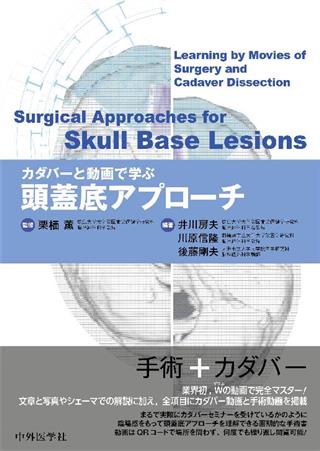
カダバーと動画で学ぶ頭蓋底アプローチ

出版社: 中外医学社
著者:
発行日: 2017-05-15
分野: 臨床医学:外科  >  脳神経外科学
ISBN: 9784498228849
電子書籍版: 2017-05-15 (1版1刷)
書籍・雑誌
≪全国送料無料でお届け≫
取寄せ目安:8~14営業日

13,200 円(税込)

電子書籍
章別単位での購入はできません
ブラウザ、アプリ閲覧

13,200 円(税込)

商品紹介

業界初、Wの動画で完全マスター! 文章と写真やシェーマでの解説に加え、全項目にカダバー動画と手術動画を掲載。まるで実際にカダバーセミナーを受けているかのように臨場感をもって頭蓋底アプローチを理解できる画期的な手術書。動画はQRコードで場所を問わず、何度でも繰り返し閲覧可能!

目次

  • カダバーと動画で学ぶ頭蓋底アプローチ

    ―目次―

    I Anterior skull base,Transnasal
     1.Transsphenoidal approach
     2.Endoscopic endonasal anterior petrosal approach
     3.Transclival approach
     4.Trans-maxillary approach
     5.Transbasal approach

    II Middle skull base
     1.Modified Dolenc approach
     2.Extradural temporopolar approach
     3.Pericavernous sinus approach
     4.Anterior petrosal approach
     5.Middle fossa approach

    III Posterior skull base
     1.Posterior-combined petrosal approach
     2.Transcondylar approach
     3.Cervicocranial approach

この書籍の参考文献

参考文献のリンクは、リンク先の都合等により正しく表示されない場合がありますので、あらかじめご了承下さい。

本参考文献は電子書籍掲載内容を元にしております。

I Anterior skull base, Transnasal

P.11 掲載の参考文献
1) Kawamata T, Ishii N, Amano K, et al. A novel simple real-time electrooculographic monitoring system during transsphenoidal surgeries to prevent postoperative extraocular motor nerve dysfunction. Neurosurg Rev. 2013;36:371-6.
2) 川俣貴一. 内視鏡下経蝶形骨手術の術中モニタリング. In:寺本明, 編. NS NOW No. 19 下垂体外科Update-大きく変わった経蝶形骨手術. 東京:メジカルビュー社;2012. p. 13-22.
3) Amano K, Hori T, Kawamata T, et al. Repair and prevention of cerebrospinal fluid leakage in transsphenoidal surgery:a sphenoid sinus mucosa technique. Neurosurg Rev. 2016;39:123-31.
4) Bergland R. Pathological considerations in pituitary tumors. Prog Neurol Surg. 1975;6:62-94.
5) Teramoto A, Sano K, Osamura R, et al. Immunohistochemical observations of the pituitary adenomas with the use of enzyme-labelled antibody method--on the residual pituitary gland and "capsule" of the adenoma. Neurol Med Chir (Tokyo). 1979;19:895-902.
6) Kawamata T, Kubo O, Hori T. Surgical removal of growth hormone-secreting pituitary adenomas with intensive microsurgical pseudocapsule resection results in complete remission of acromegaly. Neurosurg Rev. 2005;28:201-8.
7) Oldfield EH, Vortmeyer AO. Development of a histological pseudocapsule and its use as a surgical capsule in the excision of pituitary tumors. J Neurosurg. 2006;104:7-19.
8) Lee EJ, Ahn JY, Noh T, et al. Tumor tissue identification in the pseudocapsule of pituitary adenoma:should the pseudocapsule be removed for total resection of pituitary adenoma? Neurosurgery. 2009;64(3 Suppl):62-70.
9) Wrightson P. Conservative removal of small pituitary tumors:is it justified by the pathological findings? J Neurol Neurosurg Psychiatry. 1978;41:283-9.
10) Jagannathan J, Smith R, DeVroom HL, et al. Outcome of using the histological pseudocapsule as a surgical capsule in Cushing disease. J Neurosurg. 2009;111:531-9.
11) Kim EH, Ku CR, Lee EJ, et al. Extracapsular en bloc resection in pituitary adenoma surgery. Pituitary. 2015;18:397-404.
12) Xie T, Liu T, Zhang X, et al. Time to revive the value of the pseudocapsule in endoscopic endonasal transsphenoidal surgery for growth hormone adenomas. World Neurosurg. 2016;89, 65-71.
13) Kawamata T, Amano K, Hori T. Novel flexible forceps for endoscopic transsphenoidal resection of pituitary tumors:Technical report. Neurosurg Rev. 2008;31:65-8.
14) Kassam AB, Thomas A, Carrau RL, et al. Endoscopic reconstruction of the cranial base using a pedicled nasoseptal flap. Neurosurgery. 2008;63(1 Suppl 1):ONS44-52.
P.24 掲載の参考文献
1) Masuda Y, Yamamoto T, Akutsu H, et al. Usefulness of subtraction of 3D T2WIDRIVE from contrast-enhanced 3D T1WI:preoperative evaluations of the neurovascular anatomy of patients with neurovascular compression syndrome. AJNR Am J Neuroradiol. 2015;36:317-22.
2) Rivera-Serrano CM, Snyderman CH, Gardner P, et al. Nasoseptal "rescue" flap:a novel modification of the nasoseptal flap technique for pituitary surgery. Laryngoscope. 2011;121:990-3.
3) 阿久津博義. 頭蓋咽頭腫(2)内視鏡下手術. In:森田明夫, 編. 新NS NOW No. 4 脳・脊髄腫瘍摘出のための引き出し-腫瘍摘出のコツとピットフォール. 東京:メジカルビュー社;2015. p. 156-65.
4) Zanation AM, Carrau RL, Snyderman CH, et al. Nasoseptal flap takedown and reuse in revision endoscopic skull base reconstruction. Laryngoscope. 2011;121:42-6.
5) Hara T, Akutsu H, Yamamoto T, et al. Cranial base repair using suturing technique combined with a mucosal flap for CSF leakage during endoscopic endonasal surgery. World Neurosurg. 2015;84:1887-93.
6) Sakamoto N, Akutsu H, Takano S, et al. Useful 'sliding-lock-knot' technique for suturing dural patch to prevent cerebrospinal fluid leakage after extended transsphenoidal surgery--technical note--. Surg Neurol Int. 2013;4:19.
P.33 掲載の参考文献
1) Saeki N, Horiguchi K, Murai H, et al. Endoscopic endonasal pituitary and skull base surgery. Neurol Med Chir(Tokyo). 2010;50:756-64.
2) Cavallo LM, de Divitiis O, Aydin S, et al. Extended endoscopic endonasal transsphenoidal approach to the suprasellar area:anatomic considerations part 1. Neurosurgery. 2007;61(3 Suppl):24-33.
3) 堀口健太郎, 佐伯直勝. 経蝶形骨アプローチに必要な外科解剖. In:塩川芳昭, 編. NS NOW No. 15 傍鞍部病変の手術-ここの病変こそ, 解剖が手術の生命線. 東京:メジカルビュー社;2011. p. 44-53.
4) Horiguchi K, Murai H, Hasegawa Y, et al. Endoscopic endonasal skull base reconstruction using a nasal septal flap:surgical results and comparison with previous reconstructions. Neurosurg Rev. 2010;33:235-41.
5) Horiguchi K, Kato H, Nishioka H, et al. Delayed postoperative epistaxis from the posterior lateral nasal branch of the sphenopalatine artery after endoscopic endonasal approach:Case report. Interdiscip Neurosurg. 2014;1:41-3.
6) Wang J, Bidari S, Rhoton A Jr, et al. Extensions of the sphenoid sinus:a new classification. Neurosurgery. 2010;66:797-816.
7) Fernandez-Miranda JC, Prevedello DM, Madhok R, et al. Sphenoid septations and their relationship with internal carotid arteries:anatomical and radiological study. Laryngoscope. 2009;119:1893-6.
8) Mato D, Yokota H, Hirono S, et al. The vidian canal:radiological features in Japanese population and clinical implications. Neurol Med Chir(Tokyo). 2015;55:71-6.
9) Kassam AB, Prevedello DM, Thomas A, et al. Endoscopic endonasal pituitary transposition for a transdorsum sellae approach to the interpeduncular cistern. Neurosurgery. 2008;62:57-72.
10) Fernandez-Miranda JC, Gardner PA, Rastelli MM Jr, et al. Endoscopic endonasal transcavernous posterior clinoidectomy with interdural pituitary transposition. J Neurosurg. 2014;121:91-9.
11) Horiguchi K, Nishioka H, Yamada S, et al. A new multilayer reconstruction using nasal septal flap combined with fascia graft dural suturing for high-flow cerebrospinal fluid leak after endoscopic endonasal surgery. Neurosurg Rev. 2016;39:419-27.
P.50 掲載の参考文献
1) Snyderman C, Kassam A, Carrau R, et al. Acquisition of surgical skills for endonasal skull base surgery:a training program. Laryngoscope. 2007;117:699-705.
2) Dehdashti AR, Karabatsou K, Ganna A, et al. Expanded endoscopic endonasal approach for treatment of clival chordomas:early results in 12 patients. Neurosurg. 2008;63:299-309.
3) Cavallo L, Messina A, Cappabianca P, et al. Endoscopic endonasal surgery of the midline skull base:anatomical study and clinical considerations. Neurosurg Focus. 2005;19:E2.
4) Barges-Coll J, Fernandez-Miranda JC, Prevedello DM, et al. Avoiding injury to the abducens nerve during expanded endonasal endoscopic surgery:anatomic and clinical case studies. Neurosurg. 2010;67:144-54.
5) Cavallo LM, Cappabianca P, Messina A, et al. The extended endoscopic endonasal approach to the clivus and cranio-vertebral junction:anatomical study. Childs Nerv Syst. 2007;23:665-71.
6) Stippler M, Gardner PA, Snyderman CH, et al. Endoscopic endonasal approach for clival chordomas. Neurosurg. 2009;64:268-77.
7) Tomio R, Toda M, Sutiono AB, et al. Gruber's ligament as a useful landmark for the abducens nerve in the transnasal approach. J Neurosurg. 2015;122:499-503.
8) Saito K, Toda M, Tomita T, et al. Surgical results of an endoscopic endonasal approach for clival chordomas. Acta Neurochir (Wien). 2012;154:879-86.
9) Schwartz TH, Fraser JF, Brown S, et al. Endoscopic cranial base surgery:classification of operative approaches. Neurosurg. 2008;62:991-1002.
P.56 掲載の参考文献
1) Saeki N, Horiguchi K, Murai H, et al. Endoscopic endonasal pituitary and skull base surgery. Neurol Med Chir (Tokyo). 2010;50:756-64.
2) Frank G, Sciarretta V, Calbucci F, et al. The endoscopic transnasal transsphenoidal approach for the treatment of cranial base chordomas and chondrosarcomas. Neurosurgery. 2006;59(1 Suppl 1):ONS50-7.
3) 松野義晴, 堀口健太郎, 佐伯直勝, 他. 第1回千葉神経内視鏡ハンズオンセミナー~安全・低侵襲な脳神経外科手術~報告記. 千葉医学. 2007;83:53-5.
4) Cavallo LM, Esposito I, Cappabianca P, et al. Endoscopic Anatomy of the Clivus and Posterior Fossa. In:Aldo Cassol Stamm, editor. Transnasal Endoscopic Skull Base and Brain Surgery:Tips and Pearls. New York:Thieme;2011. p. 265-74.
5) Taniguchi M, Akutsu N, Kohmura E, et al. Endoscopic endonasal translacerum approach to the inferior petrous apex. J Neurosurg. 2016;124:1032-8.
6) Osawa S, Rhoton AL Jr, Seker A, et al. Microsurgical and endoscopic anatomy of the vidian canal. Neurosurgery. 2009;64:385-411.
7) Kassam AB, Prevedello DM, Thomas A, et al. Endoscopic endonasal pituitary transposition for a transdorsum sellae approach to the interpeduncular cistern. Neurosurgery. 2008;62:57-72.
8) Fernandez-Miranda JC, Gardner PA, Rastelli MM Jr, et al. Endoscopic endonasal transcavernous posterior clinoidectomy with interdural pituitary transposition. J Neurosurg. 2014;121:91-9.
9) Barges-Coll J, Fernandez-Miranda JC, Prevedello DM, et al. Avoiding injury to the abducens nerve during expanded endonasal endoscopic surgery:anatomic and clinical case studies. Neurosurgery. 2010;67:144-54.
10) Morera VA, Fernandez-Miranda JC, Prevedello DM, et al. "Far-medial" expanded endonasal approach to the inferior third of the clivus:the transcondylar and transjugular tubercle approaches. Neurosurgery. 2010;66(6 Suppl Operative):211-9.

II Middle skull base

P.101 掲載の参考文献
1) Dolenc VV. A combined epi-and subdural direct approach co carotid-ophthalmic artery aeurysms. J Neurosurg. 1985;62:667-72.
2) Dolenc VV. Anatomy and Surgery of the Cavernous Sinus. Wien, New York:Springer-Verlag;1989.
3) Dolenc VV. Transcranial epidural approach to pituitary tumors extending beyond the sellae. Neurosurgery. 1997;41:542-50.
4) Dolenc VV. Microsurgical Anatomy and Surgery of the Central Skull Base. Wien:Springer-Verlag;2003.
5) 井川房夫. 上眼窩裂外側硬膜の切開剥離を利用した手術-Sphenoparietal sinus移動のコツと注意点-. 脳神経外科速報. 2010;20:902-10.
6) 井川房夫. 脳底動脈瘤に対するtrans-sylvian approach. In:井川房夫, 他編. 前大動脈瘤・椎骨脳底動脈瘤のすべて. 大阪:メディカ出版;2016. p. 202-10.
7) 井川房夫. Dolenc approach. In:井川房夫, 他編. 頭蓋咽頭腫パーフェクトブック. 東京:中外医学社;2016. p. 62-71.
P.106 掲載の参考文献
1) Balasingam V, Noguchi A, McMenomey SO, et al. Modified osteoplastic orbitozygomatic craniotomy. Technical note. J Neurosurg. 2005;102:940-4.
2) Dolenc VV. A combined epi- and subdural direct approach to carotid-ophthalmic artery aneurysms. J Neurosurg. 1985;62:667-72.
3) 野口明男, 塩川芳昭, Delashaw JB Jr. Orbitozygomatic approachにおける顔面神経麻痺を防ぐための微小解剖. No Shinkei Geka. 2010;38:703-13.
4) Noguchi A, Balasingam V, Shiokawa Y, et al. Extradural anterior clinoidectomy. Technical note. J Neurosurg. 2005;102:945-50.
P.118 掲載の参考文献
1) Day JD, Giannotta SL, Fukushima T. Extradural temporopolar approach to lesions of the upper basilar artery and infrachiasmatic region. J Neurosurg. 1994;81:230-5.
2) Dolenc VV. A combined epi-and subdural direct approach to carotid-ophthalmic artery aneurysms. J Neurosurg. 1985;62:667-72.
3) Dolenc VV. Elements of the epidural approach to the parasellar space and adjacent regions in the central skull base. In:Dolenc VV, editor. Microsurgical Anatomy and Surgery of the Central Skull Base. Wien:Springer-Verlag;2003. p. 51-72.
4) 谷野慎, 宮原宏輔, 市原輝夫, 他. Unruptured normal sized paraclinoid aneurysm -分類と手術適応の検討, ならびにその手術手技-. 脳神経外科ジャーナル. 2016;25:236-45.
5) 森健太郎. Orbitozygomatic approachとextradural temporopolar approach-手術の工夫-. 脳神経外科ジャーナル. 2014;23:785-93.
P.126 掲載の参考文献
1) Dolenc VV. A combined epi-and subdural direct approach to carotid-ophthalmic artery aneurysms. J Neurosurg. 1985;62:667-72.
2) Dolenc VV. Elements of the epidural approach to the parasellar space and adjacent regions in the central skull base. In:Dolenc VV, editor. Microsurgical Anatomy and Surgery of the Central Skull Base. Wien:Springer-Verlag;2003. p. 51-72.
3) Day JD, Giannotta SL, Fukushima T. Extradural temporopolar approach to lesions of the upper basilar artery and infrachiasmatic region. J Neurosurg. 1994;81:230-5.
4) 森健太郎. Orbitozygomatic approachとextradural temporopolar approach-手術の工夫-. 脳神経外科ジャーナル. 2014;23:785-93.
P.136 掲載の参考文献
1) Dolenc VV. Anatomy of the cavernous sinus. In:Anatomy and Surgery of the Cavernous Sinus. New York:Springer-Verlag;1989.
2) 白馬明. 海綿静脈洞部内頚動脈瘤の直達手術. Neurosurgeons. 1989;8:222-9.
3) Fukushima T, Sameshima T. Cavernous sinus approach. In:Sameshima T, editor. Manual of Skull Base Dissection. 2nd ed. Raleigh:AF-Neurovideo;2004.
4) 鮫島哲朗, 谷川緑野, 他. 頭蓋咽頭腫に対する手術アプローチと髄液漏防止の工夫. 日本内分泌学会雑誌. 2010;86:81-2.
5) 鮫島哲朗. 海綿静脈洞の基本解剖 "Triangles". In:塩川芳昭, 担当編集. 傍鞍部病変の手術(NS NOW 15). 東京:メジカルビュー社;2011. p. 14-23.
P.152 掲載の参考文献
1) Kawase T, Toya S, Shiobara R, et al. Transpetrosal approach for aneurysms of the lower basilar artery. J Neurosurg. 1985;63:857-61.
2) Kawase T, Shiobara R, Toya S. Anterior transpetrosal-transtentorial approach for sphenopetroclival meningiomas:surgical method and results in 10 patients. Neurosurgery. 1991;28:869-75.
3) Kawase T, Shiobara R, Toya S. Middle fossa transpetrosal-transtentorial approaches for petroclival meningiomas. Selective pyramid resection and radicality. Acta Neurochir (Wien). 1994;129:113-20.
4) Kanzaki J, Kawase T, Sano K, et al. A modified extended middle cranial fossa approach for acoustic tumors. Arch Otorhinolaryngol. 1977;217:119-21.
5) Shiobara R, Ohira T, Kanzaki J, et al. A modified extended middle cranial fossa approach for acoustic nerve tumors. Results of 125 operations. J Neurosurg. 1988;68:358-65.
6) Kawase T, Bertalanffy H, Otani M, et al. Surgical approaches for vertebro-basilar trunk aneurysms located in the midline. Acta Neurochir(Wien). 1996;138:402-10.
7) Yoshida K, Kawase T. Trigeminal neurinomas extending into multiple fossae:surgical methods and review of the literature. J Neurosurg. 1999;91:202-11.
8) Fukaya R, Yoshida K, Ohira T, et al. Trigeminal schwannomas:experience with 57 cases and a review of the literature. Neurosurg Rev. 2010;34:159-71.
9) Ichimura S, Yoshida K, Sutiono AB, et al. Greater petrosal nerve schwannomasanalysis of four cases and review of the literature. Neurosurg Rev. 2010;33:477-82.
10) Schaller B, Probst R, Strebel S, et al. Trigeminocardiac reflex during surgery in the cerebellopontine angle. J Neurosurg. 1999;90:215-20.
11) Tomio R, Akiyama T, Ohira T, et al. Usefulness of facial nerve monitoring for confirmation of greater superficial petrosal nerve in anterior transpetrosal approach. Acta Neurochir(Wien). 2014;156:1847-52.
12) Ichimura S, Yoshida K, Kagami H, et al. Epidural anterior petrosectomy with subdural visualization of sphenobasal vein via the anterior transpetrosal approach--technical case report. Neurosurg Rev. 2012;35:609-13.
13) Shibao S, Toda M, Orii M, et al. Various patterns of the middle cerebral vein and preservation of venous drainage during the anterior transpetrosal approach. J Neurosurg. 2016;124:432-9.
14) Shibao S, Borghei-Razavi H, Orii M, et al. Anterior Transpetrosal Approach Combined with Partial Posterior Petrosectomy for Petroclival Meningiomas with Posterior Extension. World Neurosurg. 2015;84:574-9.
P.169 掲載の参考文献
1) Shibao S, Toda M, Orii M, et al.Various patterns of the middle cerebral vein and preservation of venous drainage during the anterior transpetrosal approach. J Neurosurg. 2016;124:432-9.
2) Amano M, Kohno M. Intraoperative continuous monitoring of evoked facial nerve electromyograms in acoustic neuroma surgery. Acta Neurochir (Wien). 2011;153:1059-67.
3) Fisch U. Transtemporal surgery of the internal auditory canal. Report of 92 cases, technique, indications and results. Adv Otorhinolaryngol. 1970;17:203-40.
4) Garcia-Ibanez E, Garcia-Ibanez JL. Middle fossa vestibular neurectomy:a report of 373 cases. Otolaryngol Head Neck Surg. 1980;88:486-90.
5) Sanna M. Hearing preservation in acoustic neuroma surgery. Middle fossa versus suboccipital approach. Am J Otol. 1987;8:500-6.
6) House WF. Surgical exposure of the internal auditory canal and its contents through the middle, cranial fossa. Laryngoscope. 1961;71:1363-85.
P.177 掲載の参考文献
1) 河野道宏. Anterior petrosal approach-工夫と注意点-. Japan J Neurosurg (Tokyo). 2012;21:845-56.
2) Sanna M. Hearing preservation in acoustic neuroma surgery. Middle fossa versus suboccipital approach. Am J Otol. 1987;8:500-6.
3) Sanna M. Surgical anatomy of the extended middle cranial fossa approach. Skull Base Surg. 1994;4:181-8.

III Posterior skull base

P.189 掲載の参考文献
1) 後藤剛夫, 大畑建治. Combined petrosal approach. In:大畑建治, 編. NS NOW No. 7 低侵襲時代の頭蓋底手術. 東京:メジカルビュー社;2009. p. 123-31.
2) 後藤剛夫, 大畑建治. Posterior petrosal approach. In:斉藤延人, 編. ビジュアル脳神経外科7 頭蓋底2:後頭蓋窩・錐体斜台部. 東京:メジカルビュー社;2012. p. 106-17.
3) Goto T, Ishibashi K, Morisako H, et al. Simple and safe exposure of the sigmoid sinus with presigmoid approach. Neurosurg Rev. 2013;36:477-82.
4) 森迫拓貴, 大畑建治. 髄膜腫摘出 神経・脳幹との剥離. In:伊達勲, 編. 新NS NOW No. 2 若手脳神経外科医が経験したい手術アプローチ. 東京:メジカルビュー社;2015. p. 104-13.
5) 森迫拓貴, 大畑建治. Posterior petrosal approach. In:森田明夫, 編. 新NS NOW No. 4 脳・脊髄腫瘍摘出のための引き出し. 東京:メジカルビュー社;2015. p. 89-109.
P.200 掲載の参考文献
1) Fukushima T, Nonaka Y, Day JD, et al. Fukushima Manual of Skull Base Dissection. 3rd ed. Raleif:AF-Neuro Video Inc;2011.
2) Tanikawa R, Sugimura T, Hino K, et al. Surgical application of skull base technique for EC-IC bypass to P2 segment. Surg Cereb Stroke(Jpn). 2006;34:440-4.
P.221 掲載の参考文献
1) 鮫島哲朗. 入門 頭蓋底手術-側頭骨アプローチのための解剖と手術の実際. 東京:メジカルビュー社;2014.
2) Sameshima T, Fukushima T. Manual of Skull Base Dissection. 2nd ed. Pittsburgh:AF-Neurovideo Inc;2004.
P.228 掲載の参考文献
1) Bertalanffy H, Seeger W. The dorsolateral, suboccipital, transcondylar approach to the lower clivus and anterior portion of the craniovertebral junction. Neurosurgery. 1991;29:815-21.
2) Seeger W. Atlas of Topographycal Anatomy of the Brain and Surrounding Structures. Wien:Springer-Verlag;1978. p. 486-9.
3) Heros RC. Lateral suboccipital approach for vertebral and vertebrobasilar artery lesions. J Neurosurg. 1986;64:559-62.
4) Perneczky A. The posterolateral approach to the foramen magnum. In:Sammi M, editor. Surgery in and around the Brain Stem and the Third Ventricle. Berlin:Springer-Verlag;1986. p. 460-89.
5) George B, Dematons C, Cophignon J. Lateral approach to the anterior portion of the foramen magnum. Application to surgical removal of 14 benign tumors:technical note. Surg Neurol. 1988;29:484-90.
6) Sen CN, Sekhar LN. An extreme lateral approach to intradural lesions of the cervical spine and foramen magnum. Neurosurgery. 1990;27:197-204.
7) Baru RP, Sekhar LN, Wright DC. Extreme lateral transcondylar approach:technical improvements and lessons learned. J Neurosurg. 1994;81:49-59.
8) Al-Mefty O, Borba LAB, Aoki N, et al. The transcondylar approach to extradural nonneoplastic lesions of the craniovertebral junction. J Neurosurg. 1996;84:1-6
9) Matsushima T, Matsukado K, Natori Y, et al. Surgery on a saccular vertebral artery-posterior inferior cerebellar artery aneurysm via the transcondylar fossa (supracondylar transjugular tubercle) approach or the transcondylar approach:surgical results and indications for using two different approaches. J Neurosurg. 2001;95:268-74.
P.236 掲載の参考文献
1) 坂田勝巳. 後頭蓋窩頭蓋底の解剖総論. In:斉藤延人, 編. ビジュアル脳神経外科7 頭蓋底2 後頭蓋窩・斜台錐体部. 東京:メジカルビュー社;2012. p. 2-15.
2) Fukushima T. Manual of Skull Base Dissection. 2nd ed. Raleigh:AF Neuro Video Inc;2004.
P.242 掲載の参考文献
1) Yamaguchi S, Eguchi K, Kiura Y, et al. Posterolateral protrusion of the vertebral artery over the posterior arch of the atlas:quantitative anatomical study using three-dimensional computed tomography angiography. J Neurosurg Spine. 2008;9:167-74.
2) Arnautovic KI, al-Mefty O, Pait TG, et al. The suboccipital cavernous sinus. J Neurosurg. 1997;86:252-62.
3) Rhoton AL Jr. The foramen magnum. Neurosurgery. 2000;47(3 Suppl):S155-93.
4) Tan M, Wang H, Wang Y, et al. Morphometric evaluation of screw fixation in atlas via posterior arch and lateral mass. Spine. 2003;28:888-95.
5) Harms J, Melcher RP. Posterior C1-C2 fusion with polyaxial screw and rod fixation. Spine. 2001;26:2467-71.
P.247 掲載の参考文献
1) 河島雅到, 松島俊夫. 頭蓋頸椎移行部の微小外科解剖. 脳神経外科ジャーナル. 2014;23:108-13.
2) 松島俊夫, 一ツ松勤, 松角宗一郎. 椎骨動脈瘤の手術と大後頭孔外側部の外科解剖. In:詠田眞治, 編. 顕微鏡下のための脳神経外科解剖XVII. 1版. 東京:サイメッドパブリケーション;2005. p. 76-86.
3) Yamaguchi S, Eguchi K, Kiura Y, et al. Posterolateral protrusion of the vertebral artery over the posterior arch of the atlas:quantitative anatomical study using three-dimensional computed tomography angiography. J Neurosurg Spine. 2008;9:167-74.
4) Rhoton AL Jr. The foramen magnum. Neurosurgery. 2000;47(3 Suppl):S155-93.

最近チェックした商品履歴

Loading...