1) 根本繁 : 新しいカテーテル素材 (高倉公朋監修 : 脳神経外科の最先端, QOLの向上のために. 先端医療技術研究所, 東京, p248-251, 1999
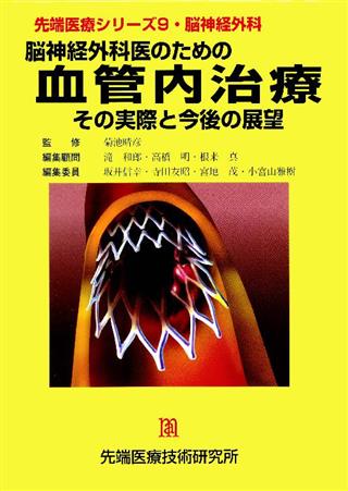
先端医療シリーズ9 脳神経外科 脳神経外科医のための血管内治療 その実際と今後の展望
商品紹介
本書の内容は、脳および脊髄領域の血管内外のすべてをカバーしており、執筆者は、それぞれの領域を現場で担当している若手脳神経外科医である。本書は、現在の日本の血管内外科の到達点、生じうる合併症を知り、将来の展望を考える上で有用であると考える。
目次
- 表紙
- カラーグラビア
- 著者一覧
- 本書を推薦する
- 本書を推薦する
- 序文
- 目次
- 第1章 機材・材料
- 1. 機材・材料
- 1.1 はじめに
- 1.2 マイクロカテーテル使用時のシステムセットアップ
- 1.3 シース(カテーテルシースイントロデューサー)
- 1.4 Yコネクター
- 1.5 ガイディングカテーテル
- 1.6 ガイドワイヤー
- 1.6.1 035inchガイドワイヤー
- 1.6.2 Exchange用ガイドワイヤー
- 1.6.3 その他のガイドワイヤー
- 1.6.4 マイクロガイドワイヤー
- 1.7 マイクロカテーテル
- 1.7.1 0ver-the-wire Microcatheters
- 1.7.2 Flow-guided Microcatheters
- 1.8 バルーンカテーテル
- 1.8.1血管閉塞用バルーンカテーテル
- 1.8.2 マイクロバルーンカテーテル
- 1.8.3 PTA(血管形成術)用バルーンカテーテル
- 2. 塞栓物質
- 2.1 はじめに
- 2.2 液体塞栓物質
- 2.2.1 シアノアクリレート系モノマー
- 2.2.2 EVAL, Eudragit-E, CAP, PVAc
- 2.2.3 95%エタノール
- 2.2.4 電気凝固による血栓
- 2.3 固形塞栓物質
- 2.3.1 PVA(polyvinyl alcohol)粒子
- 2.3.2 Pushable coil
- 2.3.3 Detachable coil
- 2.3.4 Detachable balloon
- 2.3.5 Surgical suture
- 2.3.6 Gelatin sponge (Spongel, Gelfoam),microfibrillar collagen(Avitene)
- 3. ステント
- 3.1 はじめに
- 3.2 ステントの歴史
- 3.3 ステントの分類
- 3.3.1 形状、ステントのデザインによる分類
- 3.3.2 拡張方法、delivery systemによる分類
- 3.3.3 材質による分類
- 3.4 頭頚部、脳血管に使用されているステント
- 3.4.1 Palmaz stent
- 3.4.2 EasyWall stent
- 3.4.2.1 EasyWall stentのshorteningについて
- 3.4.3 SMART stent
- 3.4.4 AVE S670 stent
- 3.5 頭頚部血管、脳血管での理想的ステント
- 4. デバイスの開発と臨床応用
- 4.1 はじめに
- 4.2 微粒子塞栓材料
- 4.3 バルーンカテーテル
- 4.4 液体塞栓材料
- 4.5 白金コイル
- 4.6 おわりに
- 第2章 周術期の管理・薬剤
- 1 はじめに
- 2 周術期の一般的管理
- 2.1 概論
- 2.2 術前準備
- 2.3 術中管理
- 2.4 術後管理
- 3 脳血管内手術における薬物治療
- 3.1 抗凝固療法・抗血小板療法
- 3.2 血栓溶解剤
- 3.3 血圧コントロール
- 3.4 鎮静・鎮痛薬、麻酔薬
- 3.5 誘発テスト、負荷テスト
- 3.6 その他
- 第3章 機能的血管解剖
- 1 はじめに
- 2 内頚動脈と椎骨・脳底動脈系の遺残血管吻合
- 3 内頚動脈系の機能的血管解剖
- 4 眼動脈と外頚動脈系の機能的血管解剖
- 5 後頭・咽頭動脈系(Occipito-Pharyngeal System)の機能的血管解剖
- 6 椎骨脳底動脈系の機能的血管解剖
- 第4章 血管内治療の病理診断
- 1 はじめに
- 2 冠動脈
- 3 大動脈ステントグラフト
- 4 脳動脈
- 5 その他の血管
- 6 おわりに
- 第5章 画像診断法の進歩
- 1. DSA
- 1.1 はじめに
- 1.2 透視装置の性能
- 1.3 装置のコントロール
- 1.4 ロードマップ機能
- 1.5 回転DSA、3D-DSA
- 1.6 ポータブルDSA
- 1.7 計測
- 1.8 主なメーカーの機種(2000年10月現在)
- 2. CT
- 2.1 はじめに
- 2.2 ルーチンスタデイ
- 2.3 ヘリカル三次元CT血管撮影(Helical 3D CT Angiography)
- 2.3.1 データ収集
- 2.3.2 三次元画像処理
- 2.3.2.1 Surface Rendering
- 2.3.2.2 Volume Rendering
- 2.3.2.3 MPR(multiplaner reformation)
- 2.3.2.4 MIP(maximal intensity projection)
- 2.3.2.5 カットモデル
- 2.3.2.6 仮想内視鏡Virtual Endoscope(VES),fly through
- 2.3.2.7 実体モデル
- 2.3.3 適応と限界
- 2.3.4 3D DSAとの対比
- 2.4 CBF study
- 2.4.1 Dynamic CT(ダイナミックCT)
- 2.4.2 Cold Xe enhanced CT(キセノンCT)
- 2.5 まとめ
- 3. MRI:とくに拡散強調画像と脳灌流画像について
- 3.1 はじめに
- 3.2 脳血管内治療の領域における従来のMRI検査の役割
- 3.3 超高速MRIの脳血管内治療領域への応用:Echo-planar imaging(EPI)法の導入
- 3.4 拡散強調画像(diffusion-weighted imaging:DWI)
- 3.4.1 拡散強調画像の原理
- 3.4.2 脳虚血におけるDWI画像の意義
- 3.4.3 DWIによる脳血管内治療に合併する脳塞栓症のスクリーニング
- 3.5 灌流画像(perfusion imaging:PI)
- 3.5.1 MRIによる脳灌流画像の原理
- 3.5.2 脳血管内治療の領域におけるPIの応用
- 3.6 おわりに
- 4. 超音波
- 4.1 はじめに
- 4.2 超音波診断機器
- 4.3 検査法
- 4.4 病変の評価
- 4.5 おわりに
- 5. 血管内脳波
- 5.1 はじめに
- 5.2 側頭葉てんかんとその焦点診断
- 5.3 海綿静脈洞内脳波記録法
- 5.4 測定例
- 5.4.1 発作間欠期脳波
- 5.4.2 発作時脳波
- 5.5 有用性・問題点・展望
- 6. SPECT
- 6.1 はじめに
- 6.2 脳血流SPECTトレーサーの特徴
- 6.2.1 123I-IMP
- 6.2.2 99mTc-ECD
- 6.3 定量評価
- 6.4 超急性期脳血管障害の脳血流SPECT
- 6.4.1 拡散強調画像所見と脳血流SPECT画像所見との比較
- 6.4.2 超急性期の治療適応
- 6.4.3 心原性脳塞栓症と脳血流SPECT画像
- 6.4.4 贅沢灌流(Luxury perfusion)の検出
- 6.5 皮質枝梗塞
- 6.6 慢性期
- 6.7 脳循環予備能の評価
- 6.8 結語
- 第6章 閉塞性疾患
- 1. 急性脳血管閉塞に対する局所再開通療法
- 1.1 はじめに
- 1.2 臨床病型の診断
- 1.3 心原性脳塞栓症
- 1.3.1適応症例の選択
- 1.3.2 閉塞部位による違い
- 1.3.3 脳血管撮影まで
- 1.3.4 脳血管撮影
- 1.3.5 血管内治療
- 1.3.6 術後管理
- 1.3.7 治療成績
- 1.3.8 NINDS studyとの関連
- 1.4 アテローム血栓性脳梗塞
- 1.4.1 Therapeutic time windowの判断
- 1.4.2 適応症例の選択
- 1.4.3 中大脳動脈閉塞症
- 1.4.4 頚部内頚動脈閉塞症
- 1.4.5 椎骨動脈起始部閉塞症
- 1.4.6 脳底動脈閉塞症
- 1.4.7 動脈原性脳塞栓症
- 1.5 まとめ
- 2. 脳静脈洞血栓症に対する血管内治療
- 2.1 脳静脈洞血栓症について
- 2.2 治療
- 2.2.1 保存的治療法
- 2.2.2 血管内治療
- 2.3 静脈洞に対するステント留置術
- 3. 頚部内頚動脈狭窄症に対する血管内治療
- 3.1 はじめに
- 3.2 適応
- 3.3 術前評価、治療手技および管理
- 3.3.1 術前評価
- 3.3.2 術前管理
- 3.3.3 治療手技および術中管理
- 3.3.4 計測
- 3.3.5 バルーンカテーテル、ステントの選択
- 3.3.6 前拡張
- 3.3.7 ステント離脱
- 3.3.8 後拡張
- 3.3.9 前拡張よりプロテクションを行う場合
- 3.3.10 術後評価
- 3.3.11 術後管理
- 3.3.12 術後検査
- 3.4 頚動脈PTA/stentingの現状と成績
- 3.5 今後の課題
- 3.5.1 狭窄部末梢での塞栓予防
- 3.5.2 フィルターデバイス(Angioguard)
- 3.5.3 総頚動脈バルーン閉塞下のPTA/stenting
- 3.6 再狭窄予防
- 3.7 結語
- 4. 椎骨動脈、鎖骨下動脈に対する血管形成術
- 4.1椎骨動脈に対する血管形成術
- 4.1.1 症状
- 4.1.2 治療の適応
- 4.1.3 方法
- 4.1.4 問題点
- 4.1.5 むすび
- 4.2 鎖骨下動脈に対する血管形成術
- 4.2.1 症状
- 4.2.2 診断
- 4.2.3 治療の適応
- 4.2.4 方法
- 4.2.5 むすび
- 第7章 血管形成術後の再狭窄
- 1. 冠動脈ステントの再狭窄
- 1.1 ステント再狭窄のメカニズム
- 1.1.1 PTCA再狭窄のメカニズム
- 1.1.2 再狭窄予防
- 1.1.3 ステント再狭窄のメカニズム
- 1.2 ステントによる再狭窄予防効果
- 1.3 血管形態、病変形態別再狭窄予防効果
- 1.3.1 血管径による再狭窄予防効果
- 1.3.2 病変の長さによる再狭窄予防効果
- 1.3.3 慢性完全閉塞病変の再狭窄予防効果
- 1.3.4 大動脈入口部
- 1.3.5 分岐部病変
- 1.4 冠動脈ステントの種類と機能
- 1.4.1 素材による分類
- 1.4.2 拡張方法による分類
- 1.4.3 形状及び製造過程による分類
- 1.4.4 付加機能による分類
- 1.5 ステント別再狭窄率
- 1.6 病変別ステント留置法
- 1.7 ステント再狭窄の形態
- 1.8 ステント再狭窄に対する対策
- 1.8.1 ステント再狭窄の予防
- 1.8.2 ステント再狭窄に対するインターベンションー再々狭窄の予防
- 2. 再狭窄に対する遺伝子治療
- 2.1 はじめに
- 2.2 血栓凝集予防療法
- 2.3 血管平滑筋細胞増殖抑制療法
- 2.3.1 細胞周期調節療法
- 2.3.1.1 アンチセンス型核酸を用いた再狭窄予防
- 2.3.1.2 デコイ型核酸を用いた再狭窄予防
- 2.3.1.3 リボザイム型核酸を用いた再狭窄予防
- 2.3.2 自殺療法
- 2.3.3 細胞内シグナル遮断療法
- 2.4 内皮再生療法(リモデリング療法)
- 2.5 おわりに
- 3. 再狭窄に対する放射線治療
- 3.1 はじめに
- 3.2 再狭窄の発症機序と放射線治療
- 3.3 血管内放射線治療に用いられる核種
- 3.4 血管内治療用器具
- 3.5 治療手技の実際
- 3.6 臨床知験の成績
- 3.6.1 冠動脈における成績
- 3.6.2 末梢血管における成績
- 3.7 被曝線量
- 3.7.1 患者被曝
- 3.7.2 術者被曝
- 3.7.3 被曝線量を減らすためには
- 3.8 血管内放射線治療の問題点
- 3.9 脳神経外科領域への応用
- 3.10 法律上の問題点と今後の展望
- 第8章 脳動脈瘤
- 1. 母血管閉塞
- 1.1 はじめに
- 1.2 balloon occlusion test
- 1.3 適応
- 1.4 周術期管理
- 1.5 治療法
- 1.6 大、巨大動脈瘤
- 1.7 解離性動脈瘤
- 1.8 症例
- 1.9 合併症とその対策
- 2. 破裂脳動脈瘤(瘤内塞栓術)
- 2.1 はじめに
- 2.2 血管内治療の利点
- 2.3 血管内治療の欠点
- 2.4 セットアップ
- 2.5 術中管理
- 2.6 手技
- 2.6.1 マイクロカテーテルの操作
- 2.6.2 coilの選択
- 2.6.3 coil挿入時の注意
- 2.6.4 終了時の判断
- 2.7 neckが大きな動脈瘤での塞栓術
- 2.8 各動脈瘤の塞栓術
- 2.8.1 前交通動脈瘤
- 2.8.2 内頚動脈瘤
- 2.8.3 中大脳動脈瘤
- 2.8.4 椎骨動脈-後下小脳動脈分岐部動脈瘤
- 2.8.5 脳底動脈瘤
- 2.8.6 上小脳動脈分岐部
- 2.9 合併症の対応
- 2.9.1 虚血の合併症
- 2.9.2 術中破裂
- 2.9.2.1 全身管理
- 2.9.2.2 血管内での対処
- 2.10 結語
- 3. 未破裂脳動脈瘤に対する血管内治療
- 3.1 はじめに
- 3.2 適応
- 3.3 方法(使用器材)
- 3.4 種々の工夫(補助手段)
- 3.5 合併症とその対策
- 3.6 今後の展望
- 4. 脳血管攣縮
- 4.1 はじめに
- 4.2 本法の適応
- 4.3 使用機器
- 4.4 周辺機器
- 4.5 方法
- 4.6 代表例
- 4.6.1 PTA列
- 4.6.2 IA例
- 4.7 結果
- 4.8 報告例
- 4.9 考察
- 4.10 まとめ
- 5. 脳動脈瘤に対する血管内治療:今後の展望
- 5.1 はじめに
- 5.2 GDCのLimitation
- 5.3 Morphological factor
- 5.4 Biological factor
- 5.5 血管内治療とTissue Engineering
- 5.6 Flow dynamic factor
- 5.7 液体塞栓物質
- 5.8 DSAからrealtime Interventional MRIへ
- 5.9 終わりに
- 第9章 血管奇形
- 1. 脳動静脈奇形塞栓術の術中治療戦略
- 1.1 はじめに
- 1.2 脳神経外科領域における血管内治療の特殊性と対策
- 1.3 脳動静脈奇形の塞栓術
- 1.4 脳動静脈奇形に対する塞栓術の適応
- 1.5 設備
- 1.6 用具
- 1.6.1 microcatheter
- 1.6.2 塞栓物質
- 1.6.2.1 固体物質
- 1.6.2.2 液体物質
- 1.6.2.3 塞栓物質の解説
- 1.7 麻酔
- 1.7.1 局所麻酔
- 1.7.2 全身麻酔
- 1.8 塞栓術中モニター
- 1.8.1 流入動脈圧(feeder pressure)
- 1.8.2 頚静脈球部酸素飽和度(Sj O2)
- 1.8.3 運動誘発電位(motor evoked potential;MEP)
- 1.9 塞栓術の実際
- 1.10 術後管理と注意点
- 1.11 治療の合併症
- 1.11.1 catheter操作による出血
- 1.11.2 正常動脈の閉塞による合併症(梗塞)
- 1.11.3 出血
- 1.12 治療のコツ
- 1.13 代表症例
- 1.14 おわりに
- 2. 脳動静脈奇形・集学的治療
- 2.1 はじめに
- 2.2 治療戦略の決定
- 2.3 根治的治療としての塞栓術
- 2.3.1 塞栓物質
- 2.3.2 塞栓方法
- 2.3.3 術後管理とfollow-up
- 2.4 Radiosurgery前の塞栓術
- 2.4.1 塞栓物質
- 2.4.2 塞栓方法
- 2.4.3 塞栓術からradiosurgeryまでのインターバル
- 2.4.4 Follow-up
- 2.5 観血的手術前の塞栓術
- 2.5.1 塞栓物質
- 2.5.2 塞栓方法
- 2.5.3 塞栓から手術までのインターバル
- 2.6 姑息的治療としての塞栓術
- 2.7 AVMに付随した動脈瘤の治療
- 2.8 まとめ
- 3. 脊髄動静脈奇形の血管内治療
- 3.1 はじめに
- 3.2 脊髄動静脈奇形の分類
- 3.3 治療戦略
- 3.4 血管内治療の実技
- 3.5 結語
- 4. 海綿静脈洞部硬膜動静脈瘻
- 4.1 病因
- 4.2 分類
- 4.3 症状
- 4.4 解剖
- 4.5 治療適応
- 4.6 治療
- 4.6.1 保存的治療
- 4.6.2 塞栓術
- 4.6.2.1 経動脈的塞栓術
- 4.6.2.2 経静脈的塞栓術
- 4.6.2.3 Anterior approach
- 4.6.2.4 Posterior approach
- 4.6.2.5 SOVの直接穿刺法
- 4.6.2.6 外科的方法
- 4.6.2.7 塞栓術の合併症
- 4.6.3 放射線治療
- 4.6.3.1 定位的放射線治療
- 5. その他の硬膜動静脈瘻
- 5.1 はじめに
- 5.2 分類
- 5.3 部位別にみた血管内治療
- 5.3.1 横・S状静脈洞部
- 5.3.1.1 分類と治療戦略
- 5.3.1.2 塞栓術
- 5.3.1.3 フォローアップ
- 5.3.2 上矢状洞部
- 5.3.2.1 治療戦略
- 5.3.2.2 塞栓術
- 5.3.2.3 経静脈的及び直接穿刺による塞栓術
- 5.3.2.4 フォローアップ
- 5.3.3 テント部
- 5.3.3.1 治療戦略
- 5.3.3.2 塞栓術
- 5.3.4 その他の部位
- 5.3.4.1 頭蓋i頸椎移行部
- 5.3.4.2 下錐体静脈洞部及びmarginal sinus
- 5.3.4.3 前頭蓋底部
- 5.4 その他の治療法
- 6. ガレン大動脈瘤およびその類似疾患
- 6.1 はじめに
- 6.2 ガレン大動脈瘤
- 6.2.1 発生学的背景および血管構築
- 6.2.2 臨床症状
- 6.2.3 治療
- 6.2.4 水頭症
- 6.3 先天性硬膜動静脈瘻
- 6.4 新生児期の血管撮影および血管内手術
- 6.4.1 新生児期の血管撮影の特異点
- 第10章 脳腫瘍
- 1. 腫瘍塞栓術
- 1.1 はじめに
- 1.2 主な対象疾患
- 1.2.1 髄膜腫meningioma
- 1.2.2 Juvenile angiofibloma
- 1.2.3 Chemodectoma
- 1.3 塞栓物質
- 1.3.1 Polyvinyl alcohol (PVA)particle
- 1.4 症例呈示
- 1.5 合併症
- 2. 悪性脳腫瘍に対する動注化学療法
- 2.1 はじめに
- 2.2 抗癌剤動注の意義
- 2.3 抗癌剤動注の適応疾患
- 2.4 術前検査および前処置
- 2.5 投与方法
- 2.6 投与薬剤
- 2.7 Blood brain barrier disruption
- 2.8 合併症
- 2.9 治療成績
- 2.10 最近行われた動注化学療法の多施設試験
- 2.11 まとめと展望
- 第11章 頭頚部疾患
- 1. 内頚動脈海綿静脈洞棲
- 1.1 はじめに
- 1.2 病因
- 1.3 症状
- 1.4 術前検査:血管造影で確認すべきポイント
- 1.5 治療
- 1.5.1 離脱式バルーンによる瘻孔閉鎖
- 1.5.2 コイルによる経動脈的または静脈的塞栓
- 1.6 合併症
- 2. 頭頚部血管病変(血管奇形、血管腫、鼻出血)の血管内治療
- 2.1 血管奇形及び血管腫の分類
- 2.2 血管腫
- 2.3 血管奇形
- 2.3.1 静脈性血管奇形(venous malformation)
- 2.3.2 毛細血管性血管奇形(capillary malfor mation)
- 2.3.3 動静脈奇形(動静脈瘻も含む)
- 2.3.4 リンパ性血管奇形(lymphatic malformation)
- 2.4 鼻出血に対する血管内治療
- 2.4.1 鼻出血(epistaxis)の分類
- 2.4.2 鼻腔の血管解剖
- 2.4.3 血管内治療
- 2.4.3.1 非外傷性の鼻出血
- 2.4.3.2 外傷性の鼻出血
- 3. 顔面けいれん
- 3.1 はじめに
- 3.2 診断
- 3.2.1 臨床症状
- 3.2.2 原因
- 3.2.3 臨床診断
- 3.2.4 画像診断
- 3.2.5 機能的診断
- 3.2.5.1方法
- 3.2.5.2 結果
- 3.3 治療
- 第12章 脳血管内治療の位置付け-特に直達手術との使い分けについて-
- 1 はじめに
- 2 脳動脈瘤
- 2.1 椎骨動脈瘤
- 2.2 脳底動脈瘤
- 2.3 内頸動脈瘤
- 2.4 その他の部位の動脈瘤
- 2.5 脳血管攣縮
- 2.6 重症クモ膜下出血例
- 2.7 高齢者における破裂脳動脈瘤
- 3 脳動静脈奇形(AVM)
- 4 閉塞性脳血管障害
- 4.1 局所再開通療法
- 4.2 血管形成術、ステント留置術
- 第13章 各種製品の臨床評価
- 1. ガンマナイフ治療の進歩
- 1.1 ガンマナイフの開発、変遷
- 1.2 ガンマナイフ治療のための画像診断-脳腫瘍
- 1.3 ガンマナイフ治療のための画像診断-脳動静脈奇形
- 1.4 集学的治療-脳動静脈奇形
- 2. 止血デバイスについて
- 2.1 はじめに
- 2.2 止血デバイスは必要か?
- 2.3 止血デバイスの種類
- 2.4 プロスター使用の実際
- 2.5 プロスターの臨床成績
- 2.6 まとめ
- 技術資料編
- OEC Mobile DSAについて
- 1 はじめに
- 2 高画質
- 3 回転陽極型X線管
- 4 大容量メモリー
- 5 おわりに
- Cutting BalloonTM
- 1 はじめに
- 2 Cutting BalloonTMの構造 / 改良
- 3 使用方法
- 4 拡張機序
- 5 Cutting BalloonTMが有用とされる病変
- 5.1 ステント内再狭窄病変
- 5.2 小血管
- 5.3 入口部病変
- 5.4 分岐部病変
- 5.5 ステント留置の前拡張
- 6 多施設における無作為割付臨床試験(REDUCE trial)
- 6.1 REDUCE1
- 6.2 REDUCE2
- 6.3 REDUCE3
- 7 今後の展望
- 血管内視鏡の現状と今後の展望
- 1 はじめに
- 2 血管内視鏡システム
- 3 血管内視鏡の手技
- 4 血管内視鏡の課題
- 4.1 カテーテル技術の課題
- 4.2 診断技術の課題
- 5 まとめ
- MRAの最近の進歩について
- 1 はじめに
- 2 MRAの撮像法
- 2.1 表示法
- 2.2 撮影法
- 2.2.1 Time-of-flight(TOF)法
- 2.2.2 Phase-contrast (PC)法
- 2.2.3 Infusion-MRA
- 最近の3D画像と血管内治療
- 1 はじめに
- 2 3次元画像処理システムRT 3D Angio Virtuoso
- 3 フルスケール・ボリューム・レンダリング・テクニック(VRT)
- PTAバルーンカテーテル 最近の進歩
- 1 はじめに
- 2 開発の流れ
- 2.1 PTAバルーン材質
- 2.2 PTAバルーンのシャフト構造
- 2.3 PTAバルーンの性能
- 3 製品の特徴
- 3.1 PTAバルーンカテーテル
- 3.2 PTCAバルーンカテーテル
- 3.3 脳血管内用バルーンカテーテル
- 4 まとめ
- Equinox Occlusion Balloon Catheterについて
- 1 はじめに
- 2 開発の経緯
- 3 特徴
- 4 使用方法
- 5 注意
- 6 おわりに
- 抗凝固剤と抗血小板剤について~アルガトロバン・塩酸チクロピジン~
- 1 アルガトロバン
- 1.1 開発の経緯
- 1.2 製品の特徴
- 1.3 今後の展望
- 2 塩酸チクロピジン
- 2.1 開発の経緯
- 2.2 製品の特徴
- 2.3 今後の展望
- テルモ社製血管造影用ガイドワイヤーについて
- 1 はじめに
- 2 開発経緯
- 3 構造
- 4 頭部領域への展開
- 5 頭部領域への新たな展開
- 6 今後の医療器開発
- DSAの血管内治療における役割と今後の展望
- 1 はじめに
- 2 DSAの歴史と原理
- 2.1 DSAの歴史
- 2.2 DSA装置の構成
- 2.3 DSA画像処理
- 3 血管内治療に有用なDSAの機能
- 3.1 透視画像処理
- 3.2 参照画像機能
- 3.3 画像解析
- 3.4 回転DSA
- 4 今後の展望
- 4.1 3次元構造の把握
- 4.2 血流動態の把握
- 5 まとめ
- 抗血栓性コーティング材料 東レ"アンスロン(R)"
- 1 はじめに
- 2 抗血栓性材料としてのアンスロン
- 2.1 ヘパリンの溶出挙動
- 2.2 ヘパリンの内部からの補給
- 2.3 アンスロンの化学構造と分子設計
- 2.4 ヘパリンの溶出速度の支配因子
- 3 アンスロン新製品の特長
- 3.1 アンギオカテーテル
- 4 おわりに
- 点滴静注用塩酸ドパミン製剤「カタボン・Low/Hi」について
- 1 はじめに
- 1.1 ドパミンの歴史
- 1.2 ドパミンの特徴
- 1.2.1 ドパミンの作用発現時間と持続時間
- 1.2.2 ドパミンの薬理作用
- 1.2.3 ドパミンの化学変化
- 2 カタボン・Low/Hiの特徴
- 2.1 開発の経緯
- 2.2 製剤的特徴
- 2.3 投与量の換算(投与量表)
- 2.4 キット製品としての有用性
- 3 カタボン・Low/Hiの効能・効果、用法・用量など
- 3.1 効能・効果
- 3.2 用法・用量
- 3.3 禁忌を含む使用上の注意など(抜粋)
- 4 カタボン・Low/Hiの臨床成績
- 5 今後の展望
- 5.1 低用量ドパミン
- 5.2 脳神経外科領域でのドパミン
- 5.3 今後の展望
- 電子走査型血管内超音波診断装置ln-Vision
- 1 はじめに
- 2 電子スキャン方式
- 3 血流のカラー表示
- 4 In-Visionの特徴
- ボストン・サイエンティフィック社マイクロカテーテルの変遷
- 1 はじめに
- 2 ボストン・サイエンティフィック社マイクロカテーテルの歴史
- 2.1 概要
- 2.2 Target Therapeutics(ターゲットセラピューティクス)社創立
- 2.3 Tracker18(トラッカー18)が開発されるまで
- 2.4 Tracker(トラッカーユニボディ、トラッカー10、 トラッカー38)
- 2.5 Fas Tracker(ファス トラッカー)
- 2.6 Fas Tracker18MX(ファス トラッカー18MX)
- 2.7 Turbo Tracker18(ターボトラッカー18)
- 2.8 Renegade18(レネゲード18)
- 2.9 Excelsior1018(エクセルシオ1018)
- 2.10 Spinnaker(スピネーカー)
- 3 今後の展望
- Detach Coil System(DCS)について
- 1 商品開発のポリシー&コンセプト
- 2 製品の特徴と効果
- 2.1 DCSデザイン
- 2.2 コイルデザイン別に見た特徴
- 3 今後の展開
- 4 まとめ
- 企業一覧
- 和文索引
- 欧文索引
- 奥付
この書籍の参考文献
参考文献のリンクは、リンク先の都合等により正しく表示されない場合がありますので、あらかじめご了承下さい。
本参考文献は電子書籍掲載内容を元にしております。
第1章 機材・材料
P.7 掲載の参考文献
P.13 掲載の参考文献
1) Taki W, Yonekawa Y, Iwata H et al : ANew Liquid Materialfor Embolization of Arteriovenous Malfonmations, AJNR 11 : 163-168, 1990
6) Brothers ME Kaufmann JCE, Fox AJ et al : n-Butyl 2-Cyanoacrylate Substitute for IBCA in Interventional Neuroradiology : Histopathologic and Polymerization Time Studies, AJNR 10 : 777-786, 1989
12) Chaloupka JC, Vinuela E Vinters HV et al : Technical feasibility and histopathologic studies of ethylene vinyl copolymer (EVAL) using a swine endovascular embolization model, AJNR 15 : 1107-1115, 1994
15) Sadato A, Taki W, Ikada Y et al : Effect of electrical thrombosis on coil embolization of experimental aneurysms, Joumal of Neurovascular Disease 2 : 235-245, 1997
16) Sadato A, Taki W, Nishi S et al : Treatment of a spontaneous carotid cavemous fistula using an electrodetachable microcoil, AJNR 14 : 334-336, 1993
17) Purdy PD, Samson D, Batjer HH et al : Preoperative Embolization of Cerebral Arteriovenous Malformations with Polyvinyl Alcohol Particles : Experience in 51 Adults, AJNR 11 : 501-510, 1990
18) Horton JA, Marano GD, Kerber CW et al : Polyvinyl alcohol foam-Gelfoam for therapeutic embolization : a synergistic mixture, AJNR 4 : 143-147, 1983
19) Halbach W, Higashida RT, Barnwell SL et al : Transarterial Platinum Coil Embolization of Carotid-Cavemous Fistulas, AJNR 12 : 429-433, 1991
21) Sadato A, Taki W, Ikada Y et al : Immediately detachable coil for aneurysm treatment, AJNR 16 : 1459-1462, 1995
22) Taki W, Handa H, Miyake H et al : New detachable balloon technique for traumatic carotid-cavemous fistulae, AJNR 6 : 961-964, 1985
23) Taki W, Handa H, Yamagata S et al : Radiopaque solidifying liquids for releasable balloon technique : a technical note, Surg Neurol 13 : 140-142, 1980
24) Benati A, Beltramello A, Colombari R et al : Preoperative embolization of arteriovenous malformations with polylene threads : techniques with wing microcatheter and pathologic results, AJNR 10 : 579-586, 1989
27) Kumar A, Kaufman SI, Patt J et al : Preoperative embolization of hypervascular head and neck neoplasms using microfibrillar collagen, AJNR 3 : 163-168, 1982
P.18 掲載の参考文献
2) Palmaz JC er al : Expandable intraluminal vascular graft, A feasibility study, Surgery 99 : 199, 1986
5) Dietrich EB, Ndlaye M, Reid DB : Stenting in the Carotid Artery ; Initial Experience in 110 Patients, J Endovasc Surg 3 : 42, 1996
6) Wholey MH, Wholey M, Bergeron P et al : Current global status of carotid artery stent placement, Cathet Cardiovas Diagn 44 : 1-6, 1998
7) 坂井信幸 : ステントによる治療, 高倉公朋監修 : 脳神経外科の最先端. 先端医療技術研究所, 東京, p92-97, 1999
12) Phatouros CC, Higashida RT, Malek AM et al : Endovascular Stenting for Carotid Artery Stenosis : Preliminary Experience Using the Shape-memory-Alloy-recoverable-technology (SMART) Stent, AJNR 21 : 732-738, 2000
P.24 掲載の参考文献
2) Hamada J, Ushio Y, Kazekawa K, Tsukahara T, Hashimoto N, Iwata H : Embolization with cellulose porous beads, I : An experimental study AJNR Am J Neuroradiol 17 : 1895-1859, 1996
3) Hamada J, Kai Y, Nagahiro S, Hashimoto N, Iwata H, Ushio Y : Embolization with cellulose porous beads, II : Clinical trial, AJNR Am J Neuroradiol 17 : 1901-6, 1996
4) Debrun G, Lacour P, Caron JP, Hurth M, Comoy J, Keravel Y : lnflatable and released balloon technique experimentation in dog-application in man, Neuroradiology 9 : 267-271, 1975
6) Taki W, Handa H, Yamagata S, Yonekawa Y, Ikada Y, Iwata H : The releasable balloon technique with activated high frequency electrical current, Surg-Neurol, 13 : 405-408, 1980
10) Taki W, Yonekawa Y, Iwata H, Uno A, Yamashita K, Amemiya H : A new liquid material for embolization of arteriovenous malformations, AJNR Am J Neuroradiol 11 : 163-168, 1990
13) Chaloupka JC, Vinuela E Vinters HV, Robert J : Technical feasibiuty and histopathologic studies of ethylene vinyl copolymer (EVAL) using a swine endovascular embolization model, AJNR Am J Neuroradiol 15 : 1107-1115, 1994
16) Spiegel SM, Vinuela E Goldwasser JM, Fox AJ, Pelz DM : Adjusting the polymerization time of isobutyl-2 cyanoacrylate, AJNR Am J Neuroradiol 7 : 109-112, 1986
18) Brothers ME Kaufmann JC, Fox AJ, Deveikis JP : n-Butyl 2-cyanoacrylate-substitute for IBCA in interventional neuroradiology : histopathologic and polymerization time studies, AJNR Am J Neuroradiol 10 : 777-786, 1989
19) 筏義人 : 生体材料学, 産業図書, 東京, p182-183, 1996
22) Sadato A, Taki W, Ikada Y, Nakahara I ; Matsumoto K, Tanaka M, Kikuchi H : Immediately detachable coil for aneurysm treatment,
23) Marks MP, Chee H, Liddell RP, Steinberg GK, Panahian N, Lane B : Amechanically detachable coil for the treatment of aneurysms and occlusion of blood vessels, AJNR Am J Neuroradiol 15 : 821-827, 1994
第2章 周術期の管理・薬剤
P.34 掲載の参考文献
1) 小笠原俊実, 竹中孝, 堀本和志, ほか : 非イオン性造影剤による血小板減少症の1例, 臨床放射線42 : 393, 1997
2) Adnan IQ, Andreas RL, Mudit S et al : Prevention and Treatment of Thromboembolic and Ischemic Complication Associated with Endovascular Procedures : Part II-Clinical Aspects and Recommendations, Neurosurgery Vol.46, No.6 : 1360-1376, 2000
3) 定藤章代, 滝和郎, 中原一郎ほか : 脳動静脈奇形の塞栓術における誘発テストの改良, 第8回日本脳神経血管内手術研究会講演集 : 43-46, 1992
4) Kobayashi S, Kageyama Y, Shimoeda N et al : Recurrence of vasospasm after endovascular vasodilating therapy in patients with subarachnoid hemorrhage, Advartce in lnterventional Neuroradiology and Intravascular Neurosurgery, Elsevier, Amsterdam, p207-210, 1996
第3章 機能的血管解剖
P.43 掲載の参考文献
1) Padget DH : The development of the cranial arteries in the human embryo, Contrib Embryol 32 : 205-261, 1948
2) Lie TA : Congenital malformations of the carotid and vertebral arterial systems, including the persistent anastomoses, in Vinken PE Bruyn GW (eds) : Handbook of Clinical Neurology, vol 12, Amsterdam, North-Holland, p289-339, 1972
4) Lasjaunias P, Berenstein A : Functional vascular anatomy of brain, spinal cord and spine, Surgical Neuroangiography, Vol 3, Springer, Berlin Heidelberg Now York Tokyo, 1990
6) Takahashi S, Suga T, Kawata Y et al : Anterior choroidal artely : angiographic analysis of variations and anomalies, AJNR 11 : 719-729, 1990
7) Abbie AA : The morphology of the fore-brain arteries, with especial reference to the evolution of the basal ganglia, J Anat 68 : 433-470, 1934
8) Komiyama M, Nishikawa M, Yasui T : Middle cerebral artery variations : duplicated and accessory arteries, AJNR 19 : 45-49, 1998
10) Lasj auni as P, Theron J : Radiological anatomy of the accessory meningeal artery, Radiology 12 : 99-104, 1976
11) Baumel JJ, Beard DY The accessory meningeal artery of man, J Anat 95 : 356-402, 1961
12) Haughton W, Rosenbaum AE : The normal and anomalous aortic arch and brachiocephalic arteries, in Newton TM, Potts DG (eds) : Radiology of the Skull and Brain, vol 2, St Louis, Mosby, p1145-1163, 1974
第4章 血管内治療の病理診断
P.51 掲載の参考文献
6) Isner JM, Baumgartner I, Ranh G et al : Treatment of thromboangiitis obliterans (Buerger’s disease) by intramuscular gene transfer of vascular endothelial growth factor : Preliminary clinical results, J Vasc Surg 28 : 964-975, 1998
8) 由谷親夫ほか : 1. ステント型人工血管挿入による血管壁の病理学的変化. b. 臨床的アプローチ. In ; 松本昭彦 (編), ステント型人工血管のすべて, 中外医学社, 東京, p44-54, 1999
9) North American Symptomatic Carotid Endoarterectomy Trial Collaborators : Beneficial effect of carotic endoarterectomy in symptomatic patients with high-grade carotid stenosis, N Engl J Med 325 : 445-453, 1991
16) Jamieson SW, Auger WR, Fedullo PF et al : Experience and results with 150 pulmonary thromboendoarterectomy operations over a 29 months period, J Thorac Cardiovasc Surg 106 : 116-127, 1993
第5章 画像診断法の進歩
P.56 掲載の参考文献
3) 久保道也, 園部眞, 杉田京一ほか : 脳血管内手術中における血管病変計測法. 臨床放射線40 : 1637-1640, 1995
4) 小林繁樹, 景山雄介, 益田一哲ほか : DSAにおける簡便かつ高精度の実測値計測ツールの開発. 第12回日本脳神経血管内手術研究会, 名古屋, 1996
P.67 掲載の参考文献
6) Anthony M, Masaryk, Richard Frayne, stal, Utility of CT Angiography and MR Angiography for the Follow-up of Experimental Aneurysms Treated with Stents or Guglielmi Detachable Coils AJNR Am J Neuroradiol 21 : 1523-1531, 2000
8) Fukasaku K, Negoro M, Fukui K et al, Three-dimensional CT angiography for embolization of aneurysm-preoperative measurement of aneurysm size and anatomical understanding in Taki W, Pickerd L, Kikuchi H eds : Advances in Interventional Neuroradiology and Intravascular Neurosurgery, Elsevier 71-74, 1996
9) Fukasaku K, Negoro M, Fukui K et al, Photomodeling and virtuan endoscop for smimulation before emboloiation uning 3D CT angiography, n Taki W, Pickerd L, Kikuchi H eds : Advances in Interventional Neuroradiology and Intravascular Neurosurgery, Elsevier 105-108, 1996
11) Touho H ; Karasawa J ; Ohnishi H, et, al, Comparison of dynamic CT and stabl exenon CT in ischemic cerebrovascular disease, AJNR Am J Neuroradiol 14 : 3, 655-60, 1990 May.
13) Thomas E, Mayer, Gerhard F Hamann, Jan Baranczyk, Dynamic CT Perfusion Imaging of Acute Stroke, JNR Am J Neuroradiol 21 : 1441-1449, 2000
14) Yonas H ; Pindzola RP ; Johnson DW ; Xenon/computed tomography cerebral blood flow and its use in clinical management, Neurosurg Clin N Am 7 : 4, 605-16, 1996 0ct.
P.74 掲載の参考文献
8) Sakai H, Sakai N, Nakahara I, et al : Embolic complication of endovascular surgery for cerebrovascular diseases : Evaluation with diffusion-weighted MR imaging, Interventional Neuroradiology (in press)
9) Bendzszus M, Koltzenburg M, Burger R et al : Silent embolism in diagnostic angiography and neurointerventional procedures : aprospective study, Lancet 354 : 1594-97, 1999
10) Lovblad KO, Pluschke W, Remonda L et al : Diffusion-weighted MRI for monitoring neurovascular interventions, Neuroradiology 42 : 134-138, 2000
14) von Kummer R, Holle R, Grzyska U et al : Interobsever agreement in assessing early CT signs of middle cerebral artery infarction, Am J Neuroradiol 17 : 1743-1748, 1996
15) Baird AE, Schlung G, Siewert B et al : Magnetic resonance perfusion-diffusion mismatch in acute human stroke : relation to ischemic lesion enlargement, Stroke 28 : 242, 1997
P.85 掲載の参考文献
1) Kimura K, Yonemura K, Terasaki T et al : Duplex carotid sonography in distinguishing acute unilateral atherothrombotic from cardioembolic carotid artery occlusion, Am J Neuroradiol 18 : 1447-1452, 1997
P.90 掲載の参考文献
6)Hahn JF, Luders H : Placement of subdural grid electrodes at the Cleveland Clinic, In : Engel J Jr, ed. Surgieal treatment of the epietpsies, New York : Raven Press, 621-7, 1987
7)Berkovic S, Andermann F, Olivier A et al : Hippocampal sclerosis in temporal lobe epilepsy demonstrated by MRI, Ann Neurol 29 : 175-82, 1981
9)Kuzniecky RI, Cascino GD, Palmini A, Jack CR Jr, Berkovic SF, Jackson GD, McCarthy G : Structural Neuroimaging, In : Engel J Jr, ed, Surgical treatment of the epielpsies, New York : Raven Press, 197-209, 1993
11) Wieser HG, Elger CE, Stodieck SR : The‘foramen ovale electrode’ : a new recording method for the preoperative evaluation of patients suffering from mesio-basal temporal lobe epilepsy, Electroencephalogy Clin Neurophysiol 61 : 314-22, 1985
12) Teramoto A, Nemoto S, Takakura K, Sasaki Y, Machida T : Selective venous sampling directly from cavemous sinus in Cushing’s syndrome, J Clin Endocrinol Metab 76 : 637-41, 1993
13) Doppman JL, Nieman LK, Chang R et al : Selective venous sampling from the cavemous sinuses is not a more reliable technique than sampling from the inferior petrosal sinuses in Cushing’s syndrome. J Clin Endocrinol Metab 80 : 2485-9, 1995
P.100 掲載の参考文献
1) 西村恒彦編, SPECT機能画像-定量化の基礎と臨床. メジカルビュー社 (東京), 1998
2) 西村恒彦編, 最新脳SPECT/PETの臨床-脳機能の検査法-. メジカルビュー社 (東京), 1995
3) Phelps ME et al : Positron emission tomography and autoradiography, In : Principles and Apphcations for The Brain and Heart, Raven Press, New York, p495, 1986
4) Iida H, Itoh I, Nakazawa H et al : Quantitative mapping of regional cerebral blood flow using iodine-123-IMP and SPECT, J Nucl Med 35 : 2019-2030, 1994
5) 飯田秀博 : IMP-ARG法. SPECT機能画像-定量化の基礎と臨床- (西村恒彦, 編). 72-78, 東京, メジカルビュー社, 1998
14) Nakagawara J, Hyogo T, Kataoka T et al : A superselective intraarterial trial using t-PA for embolic stroke : Brain Protection Grand Round, CVD grand round series, vol, 5, (Hayakawa T, et al, eds) , 169-184, Neuron Publising, Tokyo, 1998
17) 高田達郎, 中川原譲二, 瓢子敏夫ほか : 局所血栓溶解療法における脳血流量と脳梗塞出現の有無との関係 : 133Xe-SPECTによる評価. 脳卒中20 : 573-576, 1998
19) Beer HF, Blauenstein PA, Hasler PH et al : In vitro and in vivo evaluation of iodine-123-Ro 16-0154 : a new imaging agent for SPECT investigations of venzodiazepine recepters, J Nucl Med 31 : 1007, 1990
20) Hatazawa J, Satoh T, Shimosegawa E et al : Evaluation of cerebral infarction with iodine-123-iomazenil SPECT, J Nucl Med 36 : 2154, 1995
第6章 閉塞性疾患
P.111 掲載の参考文献
9) Multicentre Acute Stroke Trial-Italy (MAST-1) Group : Randomised controUed trial of streptokinase, aspirin, and combination of both in treatment of acute ischemic stroke, Lancet 346 : 1509-1514, 1995
11) The Multicenter Acute Stroke Trial-Europe Study Group : Thrombolytic therapy with streptokinase in acute ischaemic stroke, N Engl J Med 335 : 145-150, 1996
12) The National Institute of Neurological Disorders and Stroke rt-PA Stroke Study Group : Tissue plasminogen activator for acute ischemic stroke, N Engl J Med 333 : 1581-1587, 1995
15) Zeumer H, Freitag HJ, Zanella F et al : Local intraarterial fibrinolytic therapy in patients with stroke : urokinase versus recombinant tissue plasminogen activator (tPA) , Neuroradiol 35 : 159-162, 1992
P.116 掲載の参考文献
5) Dowd CF, Malek AM, Phatouros CC et al : Appllcation of a rheolytic thrombectomy deVice in the treatment of dural sinus thrombosis : anew technique, AJNR 20 : 568-570, 1999
8) Higashida RT, Helmer E, Halbach W et al : Direct thrombolytic therapy for superior saglttal smus thrombosis, AJNR 10 : S4-S6, 1989
11) Kim Sy, Suh JH : Direct endovascular thrombolytic therapy for dural sinus thrombosis : infusion of alteplase, AJNR 18 : 639-645, 1997
13) Smith TP, Higashida RT, Barnwell SL et al : Treatment of dural sinus thrombosis by urokinase infusion, AJNR 15 : 801-807, 1994
14) 高見俊宏, 鈴木俊久, 得能永夫ほか : 硬膜静脈洞血栓症, 血管内手術による血栓溶解療法の1例. 脳神経外科23 : 321-325, 1995
15) Tsai FY, Higashida RT, Matovich V et al : Acute thrombosis of the intracranial dural sinus : direct thrombolytic treatment, AJNR 13 : 1137-1141, 1992
P.124 掲載の参考文献
1) Theron JG, Courtheoux R, Alachkar F, Bouvard G, Maiza D : New tnple coaxial catheter system for carotid angiopalsty with cerebral protection, AJNR Am J Neuroradiology 11 : 869-874, 1990
3) 寺田友昭, 横手秀行, 中村善也, 津浦光晴, 中井國雄, 西口孝, 板倉徹, 林靖二, 駒井則彦, 兵谷源八, 板谷勝美 : 内頚動脈血管形成術用blocking balloon catheterの開発, 脳神経外科21 : 891-895, 1993
5) Wholey MH, Wholey M, Mathias K, Roubin GS et al : Global experience in cervical carotid artery stent placement, Cathet Cardiovasc Interv 50 : 160-167, 2000
9) 寺田友昭, 津浦光晴, 松本博之, 増尾修, 中井國雄, 板倉徹, 磯崎修二 : ガイドワイヤー先行式blocking balloon catheterを用いた内頚動脈狭窄症の血管内治療, 脳卒中の外科2000 (印刷中)
10) 寺田友昭, 津浦光晴 : 封じ込めデバイス, Heart View 4 : 1342-1346, 2000
12) Executive Committee for the Asymptomatic Carotid Atherosclerosis Study : Endarterectomy for asymptomatic carotid artery stenosis, JAMA 273 : 1421-1428, 1995
15) 岩井丈幸, 内藤功, 富沢真一郎, 福田利夫 : 内頚動脈起始部狭窄症に対するPTAにより生じた末梢塞栓に関する一剖検例での検討 : 第6回日本血管内治療学会総会抄録集 p102, 2000
16) Jordan WD, Voellinger DC, Fisher WS, Redden D, McDowell HA : A comparison of carotid angioplasty with stenting versus endarterectomy with regional anesthesia, J Vasc Surg 28 : 397-403, 1998
P.132 掲載の参考文献
3) Seoley GS, Marks MP, Dake M et al : vertebral artery stenting following percutaneous transluminal angioplasty. J Neurosurg 84 : 883-887, 1996
6) Bachmann DM, Kim RM : Transluminal dilatation for subclavian steal syndrome, AJR 135 : 995-996, 1980
8) Moran KT, Zide RS, Persson AV et al : Natural history of subclavian steal syndrome. Am Surg 54 : 643-644, 1988
11) Rodrigues-Lopes JA, Werner A, Martinez R et al : Stenting for atherosclerotic occlusive disease of the subclavian artery, Ann Vasc Surg 13 : 254-260, 1999
第7章 血管形成術後の再狭窄
P.140 掲載の参考文献
1) Rensing BJ, Hermans WR, Beatt KJ et al : Quantitative angiographic assessment of elastic recoil after percutaneous transluminal coronary angioplasty, Am J Cardiol 66 (15) : 1039-44, 1990
8) Adelman A, Cohen EA, Kimball BP et al : Acomparison of directional atherectomy with balloon angiioplasty for the lesions of the left anterior descending coronary artery, N Engl J Med 329 : 228-233, 1993
9) Hoffman R, Mintz GS, Popma JJ et al : Chronic arterial responses to stent implantation, A serial intravascular ultrasound analysis of Palmaz-Schatz stents in native coronary artenes, JACC 28 : 1134-1139, 1996
10) Fischarnan DL, Leon MB, Bain DS et al : A Randomized Of Coronary-Stent placement And BaUoon Angioplasty In The Treatment Of Coronary Artery Disease, NEJM 331 : 496-501, 1994
14) Kobayashi Y, Gregorio JD, Nobuyuki K et al : Comarison of immediate and follow-up results of the short and long NIR stent with the Palmaz-Schatz stent, Am J Cardiol 84 : 499-504, 1999
15) Mehran R, Hong MK, Lansky AJ et al : Vessel size and lesion length influence late clinical outcomes after native coronary artery stent placement, Circulation 96 : I-274 (Abstract) : 1997
16) Rubartrlli P, NIccol IL, Vema E et al : Stent Implantation Versus Balloon Angioplasty In Chronic Coronary Occlusion : Result From the GISSOC Trial : JACC 32 : 90-96, 1998
18) Simes PA, Golf Svein, Myreng Y et al : Stenting in Choronic Coronary Oocclusion (SICCO) : A Randomized, Controlled Trial of Adding Stent Implantation After Successful Angiplasty, JACC 28 : 1444-1451, 1996
22) Lefevre T, Louvard Y, Morice MC et al : Stenting of bifurcation lesions : classification, treatment, and results, CCI 49 : 274-83, 2000
23) Baim DS : ASCENT trial N evaluation of the ACS Muti-Link stent, J Invasive Cardiolol 10 Suppl B : 53B-54B, 1998 Apr
27) Yokoi H, Kimura T, Nakagawa Y et al : Long-term clinical and angiographic follow up after the Palmaz-Schatz stent restenosis (abstr. ) J am Coll Cardiol 27 (suppl A) : 224A, 1996
28) Moussa I, Di Mario C, Moses J et al : the impact of preintervention plaque area as determined by intravascular ultrasound on luminal narrowing following coronary stenting. Circulation 94 : 1528, 1996, Abstract
31) Moussa I, De Gregorio J, Di Mario C et al : The use of intravascular ultrasound and spot stenting for the treatment of long lesions and small vessels, J Invasive Cardiol 11 : 47-55, 1999 Jan
32) Galli M, Bartorelli A, Bedogni F et al : Italian BiodivYsio open registry (BiodivYsio PC-coated stent) : study of clinical outcomes of the implant of a PC-coated coronary stent, J Invasive Cardiol 12 : 452-8, 2000
33) Oberhoff MA, Cetin S, Ghobainy RA et al : Intracoronary delivery of Paclitaxel after experimental balloon angioplasty using the double balloon perfusion catheter, Circulation 102 (Supple) , II-564 : 2000
34) Sousa JE, Abizaid A, Abizaid A, Feres F et al : First human experience with Sirolimus coated BX Velocity stent : Clinical, angiographic and ultrasound late results, Circulation 102 (Supple) , II-815 : 2000
P.145 掲載の参考文献
P.151 掲載の参考文献
1) Liermartn D et al : Prophylactic endovascular radiotherapy to prevent intimal hyperplasia after stent implantation in femoropophteal arteries, Cardiovasc Intervent Radiol 17 : 12-16, 1994
6) Verin V et al : Feasibility of intracoronary β-irradiation to reduce restenosis after balloon angioplasty, A clinical pilot study, Circulation 95 : 1138-1144, 1997
7) King SB III et al : Endovascular β-radiation to reduce restenosis after coronary balloon angioplasty, Results of the beta energy restenosis trial (BERT) , Circulation 97 : 2025-2030, 1998
9) Waksman R et al : Intracoronary γ-radiation therapy after artgioplasty inhibits reccurence in patients with in-stent restenosis, Circulation 101 : 2165-2171, 2000
10) Waksman R et al : Intracoronary β-radiation therapy inhibits recurrence of in-stent restenosis, Circulation 101 : 1895-1898, 2000
11) Raizner AE et al : Inhibition of restenosis with β-emitting radiotherapy, Report of the proliferation reduction with vascular energy trial (PREVENT) , Circulation 102 : 951-958, 2000
12) Albiero R et al : Short-and intermediate-term results of P-32 radioactive β-emitting stent implantation in patients with coronary artery disease, The Milan dose-response study, Circulation 101 : 18-26, 2000
第8章 脳動脈瘤
P.160 掲載の参考文献
2) 中川原譲二 : balloon occlusion test, 菊地晴彦, 中沢省三監修 : 改訂脳神経外科領域における血管内手術法. へるす出版, 東京, p41-45, 1995
3) Brunberg JA, Frey KA, Horton JA et al : [15O]H2O positron emission tomography determination of cerebral blood flow during balloon test occlusion of the internal carotid artery, AJNR 15 : 725-732, 1994
5) 滝和郎, 菊池晴彦 : 脳動脈瘤の塞栓術, 手術手技と合併症対策, 菊地晴彦, 中沢省三監修 : 改訂脳神経外科領域における血管内手術法. へるす出版, 東京, p49-57, 1995
7) Eckard DA, O'Boynick PL, McPherson CM et al : Coil occlusion of the parent artery for treatment of symptomatic peripheral intracranial aneurysms. AJNR 21 : 137-142, 2000
8) Hacein-Bey L, Connolly ES Jr, Duong H et al : Treatment of inoperable carotid aneurysms with endovascular carotid occlusion after extracranial-intracranial bypass surgery, Neurosurgery 41 : 1225-1231 ; discussion 1231-1234, 1997
12) 安井敏裕, 岸廣成, 小宮山雅樹ほか : 破裂解離性椎骨動脈瘤に対する近位側閉塞後の再破裂機序, 脳神経外科28 : 345-349, 2000
13) 加藤徳之, 江面正幸, 高橋明ほか : 親動脈の近位閉塞が奏功せず瘤内塞栓を含めた親動脈遠位閉塞術を追加した部分血栓化動脈瘤の1例, 脳神経外科28 : 817-822, 2000
P.171 掲載の参考文献
6) 松本博之, 増尾修, 桑田俊和ほか : 動脈瘤の塞栓術でgoose neck snareによる親カテーテルの保持が有用であった1例, 脳神経外科, 25 (12) : 1127-1130, 1997
7) Kazekawa K, Kawano T, Kaneko Y et al : Percutaneous transluminal angioplasty for stenosis at the origin of the vertebral artery using snare catheters, Proceedings of the XV Symposium Neuroradiologicum, Springer, Kumamoto, 25 September-1 October,, p468-470, 1994
10) Debrun GM, Aletich VA, Kehrli P et al : Selection of cerebral aneurysms for treatment using Guglielimi detachable coils : The Preliminary University of Illinois at Chicago experience, Neurosurgery 43 : 1281-1297, 1988
12) Baxter BW, Rosso D, Lownie SP : Double microcatheter technique for detachable coil treatment of large, wide-necked intracranial aneurysms, AJNR Am J Neuroradiol 19 : 1176-1178, 1998
14) Kassel NF, Tormwe JC, Haley C, et al. : The International Cooperative Study on the timing of aneurysm surgery, Part 1 : Overall management results, J Neurosurg 73 : 18-36, 1990
P.179 掲載の参考文献
1) Debmn G, Fox A, Drake C et al : Detachable balloon and calibrated leak balloon techniques in the treatment of cerebral vascular lesions, J Neurosurg 49 : 635-649, 1978
5) Sadato A, Taki W, Ikada Y et al : Immediatry detachable coil for aneurysm treatment, AJNR 16 : 1459-1462, 1995
8) Moret J, Cognard C, Weill A et al : The“remodelling technique”in the treatment of Wide neck intracranial aneurysms, Angiographic results and clinical follow-up in 56 cases, Intervent Neuroradiol 3 : 21-35, 1997
9) Turjman E Massoud T, Ji C et al : Combined stent implantation and endosaccular coil placement for treatment of experimantal wide-necked aneurysms : a feasiblity study in swine, AJNR 15 : 1087-1090, 1994
11) Pelz DM, Lownie SP, Fox AJ : Thrombombolic events associated with the treatment of cerebral aneurysms with Guglielmi detachable coils, AJNR 19 : 1541-1547, 1998
12) Murayama Y, Vinuela E Suzuki Y et al : Development of the biologically active uglielmi detachable coil for the treatment of cerebral aneurysms, AJNR 20 : 1992-1999, 1999
P.186 掲載の参考文献
1) Anderson GB, Ashforth R, SteinDE et al : CT angiography for the detection of cerebral vasospasm in patients with acute subarachnoid hemorrhage, AJNR 21 (6) : 1011-5, 2000
2) BerUj ani GK, Bank WO, Olan WJ et al : The efficacy and safety of angioplasty for cerebral vasospasm after subarachnoid hemorrhage, Neurosurgey 42 (5) : 979-986, 1998
3) Bracad S, Aulicac S, Anxionnant R et al : Diffusion image in asymptotic focal vasospasm, Apotosis of case, J Neuroradiolo 26849 : 269-72, 1999
5) Eskridge JM, McAuliffe W, Song JK et al : Balloon angioplasty for the treatment of vasospasm : result first 50 cases, Neurosrg 42 (39) : 510-6, 1998
6) Ezura M, Takahashi A : Endovascular treatment for vasospasm, 脳と神経52 (7) : 571-9, 2000
8) 藤井芳樹, 小西善史, 佐藤栄志など : 症候性脳血管攣縮36例に対するNicardipineとpapaverineの動脈内注入療法の効果, 脳血管攣縮 9 : 429-433, 1994
12) 瓢子敏夫, 中川原譲二ほか : 脳血管攣縮に対するtransluminal angioplasy ; Diamox activated 1231-IMP-SECTによる評価, 脳血管攣縮 6 : 149-154, 1991
16) Kasell NE Helm G, Simmnons N et al : Treatment of cerebral vasospasm wit intra-arterial papaverine, J Neurosurg 77 : 848-852, 1991
17) Konishi Y, Tokitu M, Sato E et al : Percutaneous transluminal angioplasty (PTA) for vasospasm after subarachnoid hemorrhage, Cerebral Vasospasm (Ed-bySanoKetal) , Univ. of Tokyo Press Tokyo 440-442, 1990
18) 小西善史 : 脳血管攣縮に対する血管形成術-基礎的検討-杏林医学会雑誌175-182, 1993
19) 小西善史, 藤井芳樹, 佐藤栄志など : 脳血管攣縮に対する複数回のIA/PTA脳血管攣縮 13 : 213-223, 1998
20) Mayberg MR : Cerebral vasospasm, Neruosurgclin Nam 9 (3) : 615-27, 1998
21) Nemoto S : Percutaneous transluminal angioplasty, 脳と神経52 (7) : 571-9, 2000
25) 佐藤栄志, 小西善史, 前村栄冶など : SpasmにおけるTranscranial Doppler Sonography(TCD)の有用性-特にPTA療法のmonitorとして-脳血管攣縮5 : 260-264, 1990
27) 高橋明 : くも膜下出血後の脳血管攣縮に対する血管形成術, 脳神経45 : 415-430, 1993
28) Tsukahara T, Yonekawa Y, Yamamoto M et al : The endothelium-dependent response of human cerebral arteries and its change after subarachnoid hemorrhage, Cerebral vasospasm (Ed by Sano K et al) , Univ, of Tokyo Press Tokyo 227-230, 1990
30) Rordrff G, Korosheetz WJ, Coen W et al : Diffusion-weighted image in vasospasm after subarachnoid hemorrhage Stroke 308 (30) : 595-605, 1999
P.192 掲載の参考文献
1) Gughelmi G, Vinuela F, Dion J, et al : Electrothrombosis of saccular aneurysms via endovascular approach, Part 2 : Preliminary clinical experience, J Neurosurg 75 (1) : 8-14, 1991
5) Mawad ME, Mawad JK, Cartwright J, et al : Long-term histopathologic changes in canine aneurysms embolized with Guglielmi detachable coils, AJNR Am J Neuroradiol 16 (1) : 7-13, 1995
9) Murayama Y, Vinuela F, Suzuki Y, et al : Ion implantation and protein coating of detachable coils for endovascular treatment of cerebral aneurysms : concepts and preliminary results in swine models, Neurosurgery 40 (6) : 1233-43 ; discussion 1243-4, 1997
11) Raymond J, Venne D, Allas S, et al : Healing mechanisms in experimental aneurysms, I, Vascular smooth muscle cells and neointima formation, J Neuroradiol 26 (1) : 7-20, 1999
第9章 血管奇形
P.200 掲載の参考文献
5) Lasjaunias PL, Berenstein A : Surgical Neuroangiography, Berlin : Springer-Verlag, p215, 1988
6) Spetzler RF, Wilson CB, Weinstein P et al. : Normal perfusion pressure breakthrough theory, Clin. Neurosurgery 25 : 561-672, 1977
7) 平山晃康, 片山容一, 坪川孝志ほか : High-flow large arteriovenous malformationsに対する摘出術前処置としての3-0 Polyester Thread塞栓術の有用性, 第6回日本脳神経血管内手術研究会講演集, p139, 1990
8) 平山晃康, 片山容一, 坪川孝志ほか : High-flow large arteriovenous malformationsに対する術前polyester threads塞栓術前後のshunt率の検討 : 流入動脈内圧測定ならびに静脈洞酸素飽和度持続測定の意義. 第7回日本脳神経血管内手術研究会講演集, 155-161, 1991
9) 片山容一, 坪川孝志, 平山晃康ほか : polyfilament polyester threadによる塞栓術を併用した脳動静脈奇形(AW)摘出術. 脳卒中の外科21 : 487-492, 1993
10) 平山晃康, 森達郎, 川又達朗ほか : 脳動静脈奇形摘出術を安全に行うための工夫-術前血管内手術とSjO2併用の利点-集中治療8 : 1259-1260, 1996
12) HirayamaT, Katayama Y, Tsubokawa T et al : Monitoring of shunt fiow during therapeutic embolization of cerebral arteriovenous malformations with jugular bulb oxygen saturation, Neuroradiology 37 : 508-509, 1995
13) Katayarna Y, TsubokawaT, Maejima S et al : Corticospinal direct responses in humans, ldentification of the motor cortex during intracranial surgery under general anesthesia, J Neurol Neurosurg and Psychiatry 51 : 50-59, 1988
P.213 掲載の参考文献
3) Richling, B, Killer M : Endovascular management of patients with cerebral arteriovenous malfommations, in Mayberg MR, Winn HR eds : Neurosurgery Clinics of North America Vol. 11, WB Saunders, Philadelphia, p123-146, 2000
8) Aletich VA, Debrun GM : Intracranial arteriovenous malformations, The approach and technique of cyanoacrylate embolization, in Connors III JJ, Wojak JC eds : Interventional Neuroradiology, Strategies and practical techniques. WB Saunders, Philadelphia, p240-258, 1999
12) Pollock BE, Lunsford LD, Flickinger JC, Kondziolka D : The role of embolization in combination with stereotactic radiosurgery in the management of pial and dural arteriovenous malformations, in Connors III JJ, Wojak JC eds : Interventional Neuroradiology, Strategies and practical techniques. WB Saunders, Philadelphia, p267-275, 1999
15) Kiler M, Kitz K, Griffitt W et al : Radiosurgery following embolisation and/or surgery of brain AVM’s, Interventional Neuroradiology 2 : 27-33, 1996
16) Gruber A, Mazal PR, Bavinzski G et al : Repermeation of partially embolized cerebral arteriovenous malformations : a clinical, radiologic, and histologic study. AJNR 17 : 1323-1331, 1996
17) Duckwiler GR, Dion JE, Vifiuela F et al : Delayed venous occlusion following embolotherapy of vascular malfonmations in the brain, AJNR 13 : 1571-1579, 1992
19) Connors III JJ, Wojak JC : Intracranial arteriovenous malformations : general considerations, in Connors III JJ, Wojak JC eds : Interventional Neuroradiology, Strategies and practical techniques, WB Saunders, Philadelphia, p227-240, 1999
20) Vifiuela F, Dion JE, Duckwiler G et al : Combined endovascular embolization and surgery in the management of cerebral arteriovenous malfonmations : experience with 101 cases, J Neurosurg 75 : 856-864, 1991
22) Perret G, Nishioka H : Arteriovenous malformations, An analysis of 545 cases of cranio-cerebral arteriovenous malformations and fistulae reported to the cooperative study, J Neurosurg 25 : 467-490, 1996
P.218 掲載の参考文献
3) Merland JJ, Reizine D, Laurent A, Khayata MH, Casasco A, Aymard A, Gobin P : Embolization of spinal cord vascular lesions, (in) Vinuela E Halbach VV, Dion JE (eds, ) : interventional Neuroradiology, Endovascular therapy of the central nervous system, Raven Press, New York, p153-165, 1992
4) 宮本享, 永田泉, 菊池晴彦 : 脊髄動静脈奇形の手術, 脳外25 : 109-115, 1997
8) 東保肇, 唐澤淳 : 脊髄動静脈奇形の塞栓術. (in) 菊池晴彦, 中沢省三 (監修) : 脳神経外科領域における血管内手術法. へるす出版, 東京, p76-82, 1991
P.225 掲載の参考文献
3) Guo W, Pan DHC, Wu HM et al : Radiosurgery as a treatment altemative for dural arteriovenous fistulas of the cavernous sinus. AJNR 19 : 1081-1087, 1998
10) Komiyama M, Nakajima H, Nishikawa M et al : Traumatic carotid cavernous sinus fistula : Serial angiographic studies from the day of trauma. AJNR 19 : 1641-1644, 1996
11) Kuppersmith MJ, Berenstein A, Choi IS et al : Management of nontraumatic vascular shunts involving the cavemous sinus. Ophthalmology 95 : 121-130, 1988
P.240 掲載の参考文献
1) Cognard C, Houdard E, Casasco AE et al : Endovascular therapy and long-term results for intracranial dural arteriovenous fistulae, in Connord III JJ, Wojak JC eds : Interventional Neuroradiology. Strategies and Practical technique. WB Saunders Company. Philadelphia, 1999
5) 宮地茂, 岡本剛, 根来真, 吉田純 : 後頭蓋窩硬膜動静脈瘻の血管内手術に必要な血管解剖. 脳神経外科28 : 951-962, 2000
6) Halbach W, Higashida RT, Hieshima GB, Mehringer CM, Hardin CW : Transvenous embolization of dural fistulas involving the transvers and sigmoid sinuses. AJNR 10 : 385-392 1989
7) 岡本剛 : 横・S状静脈洞部硬膜動静脈瘻の血管内治療. 脳神経外科速報7 : 531-539, 1999
10) Lasjaunias P, Berenstein A : Surgical neuroangiography. Vol.1 Functional anatomy of craniofacial arteries. Berlin : Springer-Verlag, 1987
11) 桑山直也, 遠藤俊郎, 高久晃 : 頭蓋内硬膜動静脈瘻の治療. 脳外28 : 305-319, 2000
14) 新井俊成, 大野, 吉野 他 : 血栓性静脈洞炎に続発したと考えられる硬膜動静脈瘻に対して血栓融解術を試みた一例. 脳神経外科25 : 621-6, 1997
15) Croft HJ, Kallmes DF, Jensen JE, Dion JE : Percutaneous transvenous coil embolizaton of a type 4 sagittal sinus dural arteriovenous fistula : case report. Neurpsurgery 41 : 1191-4, 1997
17) 高橋明, 溝井和夫, 吉本高志 : Marginal sinus dural arteriovenous shunts. 臨床症状, 血管撮影, 治療法の検討. 脳卒中の外科20 : 384-390, 1992
20) King WA, Martin NA : Anterior fossa dural arteriovenous malformations, in Awad IA, Barrow DL eds : Dural Arteriovenous Malformations. Park Ridge, AANS, pp105-116, 1993
P.247 掲載の参考文献
2) Garcia-Monaco R, Lasjaunias P, Berenstein A : Therapeutic management of vein of Galen aneurysmal malformations, in Vinuela F, Halbach V, Dion J, eds, Interventional Neuroradiology, New York, Raven Press, p113-127, 1992
3) Gold AP, Ransohoff J, Carter S : Vein of Galenmal formation, Acta Neurol Scand 40 (S11) : 5-31, 1964
15) Zerah M, Garcia-Monaco R, Rodesch G, et al : Hydrodynamics in vein of Galen malformations, Child’s Nerv Syst 8 : 111-117, 1992
第10章 脳腫瘍
P.249 掲載の参考文献
1) Derdeyn CP, Moran CJ, Cross DT et al : Polyvinyl alcohol particle size and suspension characteristics, AJNR 16 : 1335, 1995
3) Manelfe C, Lasjaunias P, Ruscalleda J : Preoperative embolization of intracranial meningiomas. A JNR 7 : 963, 1986
P.253 掲載の参考文献
2) Chamberlain MC, Kormanik PA : Practical guidelines for the treatment of malignant gliomas, West J Med 168 : 114-120, 1998
3) Pechl V, Peterson K, Caimcross J G : Chemotherapy for brain tumors, Oncology (Huntingt) 12 : 537-543, 1998
4) Hosh P, Sappino AP, Tribolet Nd et al : Malignant glioma : should chemotherapy be overthrown by experimental treatments? Ann Oncol 9 : 589-600, 1998
9) 兵頭明夫, 吉井与志彦, 樋口修ほか : 悪性脳腫瘍に対する超選択的動注化学療法, 第6回日本脳神経血管内手術研究会講演集 267-273, 1991
13) Chow KL, Gobin YP, Cloughesy T et al : Prognostic factors in recurrent glioblastoma multiforme and anaplastic astrocytoma treated with selective intraarterial chemotherapy, Am J Neuroradiol 21 : 471-478, 2000
14) 吉井与志彦, 兵頭明夫, 松丸祐司ほか : 超選択的動注療法における抗癌剤の分布, 腫瘍内濃度, CT研究17 : 125-131, 1995
15) Theron J, Villemure JG, Worthington C et al : Superselective intracerebral chemotherapy of malignant tumours with BCNU. Neuroradiological considerations, Neuroradiology 28 : 118-125, 1986
17) 兵頭明夫 : 悪性脳腫瘍に対する超選択的動注化学療法, 菊池晴彦, 中沢省三編 : 改訂脳神経外科領域における血管内手術法. へるす出版, 東京, p.194-199, 1999
19) 松丸祐司, 兵頭明夫, 吉井与志彦ほか : 悪性脳腫瘍に対する超選択的動注化学療法, 合併症の検討を中心に, 第8回日本脳神経血管内手術研究会講演集59-63, 1993
23) Safdari H, Mompeon B, Dubois JB et al : Intraarterial 1,3-bis (2-chloroethyl) -1-nitrosourea chemotherapy for the treatment of malignant gliomas of the brain : a preliminary report, Surg Neurol 24 : 490-497, 1985
25) Feun LG, Wallace S, Yung WK et al : Phase-Itrial of intracarotid BCNU and cisplatin in patients with malignant intracerebral tumors, Cancer Drug Deliv 1 : 239-245, 1984
27) Tyler JL, Yamamoto YL, Diksic M et al : Pharmacokinetics of superselective intra-arterial and intravenous[11C]BCNU evaluated by PET. J Nucl Med 27 : 775-780, 1986
28)山崎俊樹, 長尾聖一, 加川隆登ほか : 動脈内投与いよるMCNUの血中濃度と脳腫瘍内濃度の検討, 脳外23 : 963-969, 1995
32) Miyagami M, Kagawa Y, Tsubokawa T : ACNU delivery to malignant glioma tissue by osmotic blood brain barrier modification with intracarotid infusion of hyperosmoral mannitol No Shinkei Geka 13 : 955-963, 1985
33) 渡辺正人, 武田憲夫, 田中隆一 : 大脳悪性神経膠腫に対するRanimustatine(MCNU)の動注, 局注療法の経験, 癌と化学療法22 : 811-814, 1995
38) Roman-Goldstein S, Mitchell P, Crossen JR et al : MR and cognitive testing of patients undergoing osmotic blood-brain barrier disruption with intraarterial chemotherapy, Am J Neuroradiol 16 : 543-553, 1995
43) Tfayli A, Hentschel P, Madajewicz S et al : Toxicities related to intraarterial infusion of cisplatin and etoposide in patients with brain tumors, J Neurooncol 42 : 73-77, 1999
44) Kleinschmidt-DeMasters BK, Geier JM : Pathology of high-dose intraarterial BCNU, Surg Neurol 31 : 435-443, 1989
45) Mansfield SH, Castillo M : MR of cis-platinum-induced optic neuritis, Am J Neuroradiol 15 : 1178-1180, 1994
48) Neuwelt EA, Brummett RE, Doolittle ND et al : First evidence of otoprotection against carboplatin-ilduced hearing loss with a two-compartment system in patients with central nervous system malignancy using sodium thiosulfate. J Pharmacol Exp Ther 286 : 77-84, 1998
50) Fortin D, McAllister LD, Nesbit G et al : Unusual cervical spinal cord toxicity associated with intraarterial carboplatin, intraarterial or intravenous etoposide phosphate, and intravenous cyclophosphamide in conjunction with osmotic blood brain-barrier disruption in the vertebral artery, Am J Neuroradiol 20 : 1794-1802, 1999
51) Shapiro WR, Green SB : Reevaluating the efficacy of intra-arterial BCNU, J Neurosurg 66 : 313-315, 1987
第11章 頭頚部疾患
P.260 掲載の参考文献
7) Halbach VV et al : Transvenous embolization of direct carotid cavemous fistulas. AJNR 9 : 741-747, 1988
8) Halbach VV et al : Transarterial platinum coil embolization of carotid-cavernous fistulas, AJNR 12 : 429-433, 1991
11) 岡田雅彦 : 外傷性およびsingle orificeの頚動脈海綿静脈洞瘻の塞栓術, 脳神経外科領域における血管内手術法. へるす出版, p98-104, 1991
P.266 掲載の参考文献
3) Komiyania M, Nishikawa M, Kan M et al : Endovascular treatment of intractable oronasal bleeding associated with severe craniofacial injury, J Trauma 44 : 330-334, 1998
7) Merland JJ, Riche MC, Hadjean E et al : The use of superselective angiography, embolization, and surgery in the current management of cervicocephalic vascular malformations (350 cases) . Symposium on vascular malformations and melanotic lesions, in Williams HB eds, CV Mosby, St. Louis, p135-143, 1983
13) 辻口幸之助, 今井啓介, 戸田千綾ほか : 頭頸部顔面領域におけるvenous malformationの治療法. エタノール注入を中心として. 形成外科 41 : 133-138, 1999
P.271 掲載の参考文献
1) Yainashita K, Hojo M, Okamoto S et al : Possible role of neurointerventional techniques in the diagnosis of hemifacial spasm, AJNR Am J Neuroradiol 18 : 287-290, 1997
4) Moller AR : The cranial nerve vascular compression syndrome : II. A review of pathophysiology, Acta Neurochir (Wien) 113 : 24-30, 1991
6) Jannetta PJ : Tupical or atypical hemifacial spasm, J Neurosurg 89 : 346-347, 1998
9) Blair RL, Berry H : Spontaneous facial movement. J Otolaryngol 10 : 459-462
12) 岡村知實, 青木秀夫, 柳井和年, ほか : 脳神経 (V, VII-XI) 起始部のX線撮影上の位置.神経血管減圧術に関連して. Neurol Med Chir (Tokyo) 23 : 776-782, 1983
第13章 各種製品の臨床評価
P.282 掲載の参考文献
1) Leksell L : Cerebral radiosurgery, I, Gammathalamotomy in two cases of intractable pain, Acta Chir Scand 134 : 585-95, 1968
3) Leksel L, Backlund EO and Johansson L : Treatment of craniopharyngiomas, Acta Chir Scand 133 : 345-50, 1967
4) Steiner L, Leksell L, Greitz T et al : Stereotaxic radiosurgery for cerebral arteriovenous malfommations, Report of a case, Acta Chir Scand 138 : 459-64, 1972
5) Duma CM, Jacques D and Kopyov OV : The treatment of movement disorders using Garnma Knife stereotactic radiosurgery, Neurosurg Clin N Am 10 : 379-89, 1999
8) Regis J, Bartolomei F, Metellus P et al : Radiosurgery for trigeminal neuralgia and epilepsy, Neurosurg Clin N Am 10 : 359-77, 1999
P.286 掲載の参考文献
1) A. R. Gruentzig : Transluminal dilatation of coronary artery stenosis, Lancet 1 : 263, 1978
2) TNoguchi, S Miyazaki, S Yasuda, K Baba, H Sumida, S Daikoku, Y Goto, and H Nonogi : Arandomized controlled trial of Prostar Plus for hemostasis in patients after coronary angioplasty, European Journal of Vascular and Endovascular Surgery 19 : 451-455, 2000
技術資料編
P.293 掲載の参考文献
2) Barath P : Microsurgical Dilatation Concept : Animal Data, J Invas Cardiol 8 Suppl A : 2A-5A, 1996
4) Chin W : Effect of multiple inflations in cutting balloon angioplasty : Comparison with single inflation (ab-stract) , Jpn J Circ 61 (Suppl) : 432, 1997
5) 末吉浩一郎 : Cutting Balloonの初期成績-QCAとIVUSによる検討-, 心血管インターベンション Vol.11 No.2 : 155-160, 1996
6) 高野雅充 : Cutting Baloon Angioplastyにおける血管内エコーの有用性, 冠疾患誌4 : 123-127, 1998
8) 塚原玲子 : 血管内超音波より評価したカッティングバルーンの急性期拡張および遠隔期再狭窄の機序, 心血管インターベンション Vol.11 No.5 : 439-442, 1996
9) 木下順久 : 冠動脈ステント内再狭窄病変に対するcutting balloon angioplastyの初期および遠隔期成績-Plain old balloon angioplastyとの比較-, 心血管インターベンション Vol.13 No.5 : 409-414, 1998
10) Albiero R : Cutting Balloon Angioplasty for the Treatment of In-Stent Restenosis, Cathet Cardiovasc Interv 50 : 452-459, 2000
11) Ergene O : Comparison of Angiographic and Clinical Outcome after Cutting Balloon and Conventional Balloon Angioplasty in Vessels Smaller than 3mm in Diameter : A Randomized Trial, J Invas Cardiol Vol.10 No.2 : 70-75, 1998
12) Muramatsu T : Efficacy of Cutting Balloon Angioplasty for Lesions at the Ostium of the Coronary Arteries, J Invas Cardiol Vol.11 No.4 : 201-206, 1999
13) Nakamura M : Cutting Balloon Angioplasty Priorto Stentingisa Useful Strategy for the Reduction of Stent Restenosis (abstract) , Am J Cardiol Vol.84 (Suppl 6A) : 58, 1999
P.296 掲載の参考文献
1) 宮本享, 菊池晴彦ほか : 頭蓋外脳血管内視鏡の応用, 脳神経外科 17 (7) : 625-628, 1989
2) 岡本剛, 宮地茂, 根来真ほか : 血管内視鏡, CLINICAL NEUROSCIENCE 17 (6) : 649-652, 1999
3) 酒井俊太, 水野杏一 : 冠血管内視鏡, Heart View 2 (12) : 1320-1325, 1998
4) 内田康美, 金井正仁 : 血管内視鏡と血管内エコーによる急性冠症候群の予知, Coronary 15 (3) : 157-164, 1998
P.306 掲載の参考文献
1) O Gurvit, M Skalej, R Siekmann, U Ememann, K Voigt : Rotational Angiography and 3D Reconstruction in Neuroradiology, electro medica 68 (1) : 31, 2000
P.310 掲載の参考文献
2) Gruntzig A : Percutane Rekanalisation chronischer arterieller Verschiusse mit einm neuen Dilatationskatheter, Dtsch Med Wochenschr 99 : 2502-2510, 1974
P.317 掲載の参考文献
1) 岡田和悟, 足立智英, 小林祥泰 : 特集 脳梗塞急性期の治療 急性期の薬物療法 3) 抗トロンビン薬 (argatroban) の適応と使い方, Geriatric Medicine 36 : 694-700, 1998
2) 松尾武文, 小出昌伸, 松尾美也子 : 特集透析療法における抗凝固薬の見直し 3合成抗トロンビン薬-アルガトロバン, メシル酸ナファモスタット-, 透析フロンティア 7 : 9-14, 1997
3) 岩井武尚, 数井秀器, 櫻井恒久, 矢野孝 : 今月の話題 慢性動脈閉塞症重症例の治療 up to date, Ther Res 19 : 3637-3653, 1998
4) 中島光好, 梅村和夫, 川井洋 : 脳血栓症における選択的抗トロンビン剤アルガトロバンの効果, 医学と薬学 35 : 1061-1071, 1996
5) 須賀操, 幸敏志, 杉本次郎, 中島光好 : 光増感反応を利用した中大脳動脈閉塞血栓症モデルにおける選択的抗トロンビン剤アルガトロバンとトロンボキサンA2合成酵素阻害剤オザグレルナトリウムの薬効比較, 新薬と臨床 45 : 1925-1933, 1996
6) Yamashita T et al : The regional cerebral blood flow amelioration of argatroban in the acute stage of cerebral thrombosis, The Keio J Med 49 (supple) : A141-A144, 2000
8) Akiguchi I et al : Effects of arttithrombin on Binswanger’s disease with antiphospholipid antibody syndrome, Neurology 52 : 398-401, 1999
9) Donovan FM et al : Thrombin induces apotosis in cultured neurons and astrocytes via a pathway requiring tyrosine kinase and RhoA actiVities, J Neurosci 17 : 5316-5326, 1997
P.321 掲載の参考文献
1) Takayasu K, Murayama Y, Moriyama N et al : Plasticcated guide wire for hepatic arteriography. Radiology 173 : 580, 1989
3) 鈴木孝治, 川村研二, 津川龍三ほか : 尿路におけるテルモ社製ガイドワイヤーの使用経験 : 泌尿記要 33 : 638-643, 1987
4) 久呉高博, 村田幸彦, 田辺進ニラジフォーカス・ガイドワイヤーM : INTERVENTIONAL RADIOLOGY OCT VOL.10 NO.4 : 402-405, 1995
5) 鈴木雄一 : 実用形状記憶合金, 工業調査会, 東京, P46~51, 1987
P.325 掲載の参考文献
1) 松岡昭治 : 経静脈性血管造影法. 医歯薬出版株式会社, 東京, p3-55, 1994
2) R Anxionnat, S Bracard, J Macho, E Da Costa, R Vaiuant, L Launmay, Y Trousset, R Romeas and L Picard : 3D Angiography Clinical interest First applications in interventional neuroradiology, J Neuroradiology, 25 (4) : 251-262, 1998
3) 藤井千蔵 : 近未来のデジタルX線システム・X線平面検出器の現状と将来. 全国循環器撮影研究会誌, Vol.12, 2000
P.332 掲載の参考文献
2) 太田宗夫ほか, 各種ショックに対する新しい塩酸ドパミン複合製剤“カタボン”の使用感, 治療 73 (3) : 883-888, 1991
4) Bellomo R et al : Hemodynamic support and the role of dopamine, Kidney Int : 53 (suppl 66) : s71-s74, 1998
7) 阿部聡ほか : 破裂脳動脈瘤術後のHypertension-Hypervolemia療法で発生する重大なPitfall, 脳神経外科 20 (4) : 357-365, 1992
P.336 掲載の参考文献
1) 石沢錠二ほか : 頚動脈病変のステント留置における超音波診断の意義, 脳神経外科速報 9 (12) : 1023-1029, 1999
2) 中村貢ほか : 血管内超音波による画像診断 : 内頚動脈解離に対する応用, Neurosonology 10 (3) : 123-128, 1997
3) 西田幸生ほか : ChromaFloによるステント内腔面積測定の有用性, Jpn Circ J Vol63 Suppl I : 487, 1999
4) E I Cespedes et al : Angiographically undetected stent malapposition resolved by intravascular ultrasound and flow imaging. J Vascular Investigation 4 (2) : 81-84, 1998
5) 大塚雅人ほか : 血管内超音波イメージングPTCAカテーテル (オラクルメガソニック) を用いたPTCAの初期成績, Jpn J Interv Cardiol 14 (I) : 143, 1999
6) 加藤健一ほか : Balloon付きICUSを初回拡張に使用したステント植込み術の初期成績-連続例におけるコスト削減の試み-, Jpn J Interv Cardiol 14 (I) : 166, 1999
P.342 掲載の参考文献
1) 兵頭明夫, 根本繁 : GDCを用いた脳動脈瘤血管内治療. 医学書院, 東京, p11-12, 1999
P.344 掲載の参考文献
1) Daniel A Rufenacht, 杉生憲志 : Detach Coil System (DCS : William COOK Europe) の脳血領域における有用性, HIT No.17 ; 13-19, 1999
2) Philippe Gailloud, Daniel A Rufenacht et al : In Vitro Models of Intracranial Arterovenous Fistulas for the Evaluation of New Endovascular Treatment Materials, AJNR Am J Neuroradiol February 20 : 291-295, 1999
3) Michel Piotin, Daniel A Rufenacht et al : Dense Packing of Cerebral Aneurysms, An In Vitro Study with Detachable Platinum Coils, AJNR Am J Neuroradiol April 21 : 757-760, 2000
最近チェックした商品履歴
Loading...
販売価格 770 円(税込)
第1章 機材・材料
-
第1章 機材・材料P.1
-
1. 機材・材料P.1
-
1.1 はじめにP.1
-
1.2 マイクロカテーテル使用時のシステムセットアップP.1
第2章 周術期の管理・薬剤
-
第2章 周術期の管理・薬剤P.26
-
1 はじめにP.26
-
2 周術期の一般的管理P.26
-
2.1 概論P.26
第3章 機能的血管解剖
-
第3章 機能的血管解剖P.35
-
1 はじめにP.35
-
2 内頚動脈と椎骨・脳底動脈系の遺残血管吻合P.35
-
3 内頚動脈系の機能的血管解剖P.36
第4章 血管内治療の病理診断
-
第4章 血管内治療の病理診断P.44
-
1 はじめにP.44
-
2 冠動脈P.44
-
3 大動脈ステントグラフトP.47
第5章 画像診断法の進歩
-
第5章 画像診断法の進歩P.52
-
1. DSAP.52
-
1.1 はじめにP.52
-
1.2 透視装置の性能P.52
第6章 閉塞性疾患
-
第6章 閉塞性疾患P.102
-
1. 急性脳血管閉塞に対する局所再開通療法P.102
-
1.1 はじめにP.102
-
1.2 臨床病型の診断P.102
第7章 血管形成術後の再狭窄
-
第7章 血管形成術後の再狭窄P.133
-
1. 冠動脈ステントの再狭窄P.133
-
1.1 ステント再狭窄のメカニズムP.133
-
1.1.1 PTCA再狭窄のメカニズムP.133
第8章 脳動脈瘤
-
第8章 脳動脈瘤P.153
-
1. 母血管閉塞P.153
-
1.1 はじめにP.153
-
1.2 balloon occlusion testP.153
第9章 血管奇形
-
第9章 血管奇形P.193
-
1. 脳動静脈奇形塞栓術の術中治療戦略P.193
-
1.1 はじめにP.193
-
1.2 脳神経外科領域における血管内治療の特殊性と対策P.193
第10章 脳腫瘍
-
第10章 脳腫瘍P.248
-
1. 腫瘍塞栓術P.248
-
1.1 はじめにP.248
-
1.2 主な対象疾患P.248
第11章 頭頚部疾患
-
第11章 頭頚部疾患P.256
-
1. 内頚動脈海綿静脈洞棲P.256
-
1.1 はじめにP.256
-
1.2 病因P.256
第12章 脳血管内治療の位置付け-特に直達手術との使い分けについて-
-
第12章 脳血管内治療の位置付け-特に直達手術との使い分けについて-P.272
-
1 はじめにP.272
-
2 脳動脈瘤P.272
-
2.1 椎骨動脈瘤P.273
第13章 各種製品の臨床評価
-
第13章 各種製品の臨床評価P.278
-
1. ガンマナイフ治療の進歩P.278
-
1.1 ガンマナイフの開発、変遷P.278
-
1.2 ガンマナイフ治療のための画像診断-脳腫瘍P.278