1) 岡山昭彦. 身体診察のすすめ. 宮崎県内科医会誌 2006;69.
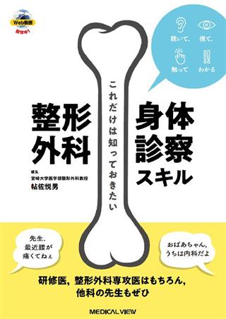
これだけは知っておきたい 整形外科身体診察スキル
商品紹介
月刊「関節外科」の連載記事「これだけは知っておきたい整形外科的徒手検査法」をベースに,整形外科の身体診察をイチから学びたい人のために,診察の基礎から画像を含めた各種検査法,診断後の治療の流れまでを各部位ごとに解説。
若手整形外科医だけではなく,研修医や総合診療医も使えるように,専門用語,重要な知識などを「Tips」として欄外に記載し,徒手検査法にはWeb動画を掲載。
目次
- I これだけは知っておきたい 身体診察を始める前の注意点
身体診察の重要性
上手な医療面接とは?
身体診察のコツ・注意点
II これだけは知っておきたい 身体診察の基本
視る:視診の基本
聴く:問診の基本
触る:触診ほかの基本
画像検査の基本
III これだけは知っておきたい 上肢の身体診察
肩関節の診察
肘関節・手関節・手指の診察
IV これだけは知っておきたい 脊椎・体幹・骨盤の身体診察
頚部・上部体幹の診察
腰背部・骨盤の診察
V これだけは知っておきたい 下肢の身体診察
股関節の診察
膝関節の診察
足関節・足部の診察
VI これだけは知っておきたい 身体診察後の対応
カルテの書き方
インフォームド・コンセントの実際
コンサルトの方法
リハビリテーション処方
必ず知っておきたい医療安全の知識
この書籍の参考文献
参考文献のリンクは、リンク先の都合等により正しく表示されない場合がありますので、あらかじめご了承下さい。
本参考文献は電子書籍掲載内容を元にしております。
I これだけは知っておきたい 身体診察を始める前の注意点
P.4 掲載の参考文献
P.7 掲載の参考文献
1) 斎藤清二. 医師と患者のコミニュニケーション. 東京:日医総研;2005.
2) 向原 圭. 医療面接 根拠に基づいたアプローチ. 東京:文光堂;2006.
P.13 掲載の参考文献
1) 日本整形外科学会, 日本リハビリテーション医学会. 関節可動域表示ならびに測定法(平成7年2月改訂). 日本整形外科学会雑誌 1995;69:240-50.
2) 日本整形外科学会 編. 日本整形外科学会評価基準・ガイドライン・マニュアル集 第3版. 1999.
3) 日本手外科学会. 手の機能評価表 第4版. 2006.
II これだけは知っておきたい 身体診察の基本
P.31 掲載の参考文献
1) 中村利孝, 松野丈夫 監. 標準整形外科学 第13版. 東京:医学書院;2017, 122-56.
2) 星野雄一, 吉川秀樹, 斉藤和行 編. NEW エッセンシャル 整形外科学. 東京:医歯薬出版;2012, 47-70.
3) Hoppenfeld JD, Hoppenfeld S. 整形外科医のための神経学図説原書第2版. 東京:南江堂;2019, 2-5.
4) 日本整形外科学会, 日本リハビリテーション医学会. 関節可動域表示ならびに測定法. 日整会誌 1995;69:240-50.
III これだけは知っておきたい 上肢の身体診察
P.71 掲載の参考文献
1) Halder AM, Zhao KD, Odriscoll SW, et al. Dynamic contributions to superior shoulder stability. J Orthop Res 2001;19:206-12.
2) Warner JJ, Deng XH, Warren RF, et al. Static capsuloligamentous restraints to superior-inferior translations of the glenohumeral joint. Am J Sports Med 1992;20:675-85.
3) O'Connell PW, Nuber GW, Mileski RA, et al. The contribution of the glenohumeral ligaments to anterior stability of the shoulder. Am J Sports Med 1990;579-84.
4) Debski RE, Wong EK, Woo SL, et al. In situ force distribution in the glenohumeral joint capsule during anterior-posterior loading. J Orthop Res 1999;17:769-76.
5) Inman VT, Saunders JB, Abbott LC. Observations on the function of the shoulder joint. J Bone Joint Surg 1944;26:1-30.
7) 米田 稔. 肩関節鏡下手術 第1版. 東京:文光堂;2010.
8) 中村利孝, 松野丈夫 監. 標準整形外科学 第13版. 東京:医学書院;2017.
9) 坂井建雄, 松村譲兒 監訳. プロメテウス解剖学アトラス 第2版. 東京:医学書院;2011.
10) 西中直也. 肩. 関節外科 2014;33(4月増):12-23.
11) 幸原伸夫 訳. 末梢神経と筋のみかた 原著第5版. 東京:診断と治療社;2016.
12) Jobe FW, Moynes DR. Delineation of diagnostic criteria and a rehabilitation program for rotator cuff injuries. Am J Sports Med 1982;10:336-9.
13) Lee CK, Itoi E, Kim SJ, et al. Comparison of muscle activity in the empty-can and full-can testing positions using 18F-FDG PET/CT. J Orthop Surg Res 2014;9:85.
14) Mochizuki T, Sugaya H, Uomizu M, et al. Humeral insertion of the supraspinatus and infraspinatus. New anatomical findings regarding the footprint of the rotator cuff. J Bone Joint Surg Am 2008;90:962-9.
15) Hertel R, Ballmer FT, Lombert SM, et al. Lag signs in the diagnosis of rotator cuff rupture. J Shoulder Elbow Surg 1996;5:307-13.
16) Hughes PC, Taylor NF, Green RA. Most clinical tests cannot accurately diagnose rotator cuff pathology:a systematic review. Aust J Physiother 2008;54:159-70.
17) Hegedus EJ, Goode A, Campbell S, et al. Physical examination tests of the shoulder:a systematic review with meta-analysis of individual tests. Br J Sports Med 2008;42:80-92.
18) Rockwood CA, Jr, et al, editors. The Shoulder. 4th ed. Philadelphia:W. B. Saunders;2009.
19) Walch G, Boulahia A, Calderone S, et al. The 'dropping' and 'hornblower's' signs in evaluation of rotator-cuff tears. J Bone Joint Surg Br 1998;80:624-8.
20) Sgroi M, Loitsch T, Reichel H, et al. Diagnostic value of clinical tests for infraspinatus tendon tears. Arthroscopy 2019;35:1339-47.
21) Gerber C, Hersche O, Farron A. Isolated rupture of the subscapularis tendon. J Bone Joint Surg Am 1996;78:1015-23.
22) Barth JRH, Burkhart SS, De Bee JF. The Bear-Hug Test:A new sensitive test for diagnosing a subscapularis tear. Arthroscopy 2006;22:1076-84.
23) Burkhart SS, Brady PC. Arthroscopic subscaplaris repair:Surgical tips and pearls A to Z. Arthroscopy 2006;22:1014-27.
24) 石田康行, 帖佐悦男, 矢野浩明, ほか. 肩甲下筋負荷テストと鏡視所見の検討. 肩関節 2010;34:609-11.
25) Gerber C, Hersche O, Farron A. Isolated rupture of the tendon of the subscaplaris muscle. Clinical features in 16 cases. J Bone Joint Surg Br 1991;73:389-94.
26) 宮武克年, 武田芳嗣, 藤井幸治 ほか. 肩甲下筋腱断裂に対する術前徒手テストとMRIの診断率. 関節外科 2015;34:968-76.
27) Burkhart SS, Tehrany AM. Arthroscopic subscapularis tendon repair:Technique and preliminary results. Arthroscopy 2002;18:454-63.
28) Scheibel M, Habermeyer P. Subscapularis dysfunction following anterior surgical approaches to the shoulder. J Shoulder Elbow Surg 2008;17:671-83.
29) Bartsch M, Greiner S, Haas NP, et al. Diagnostic values of clinical tests for subscapularis lesions. Knee Surg Sports Traumatol Arthrosc 2010;18:1712-7.
30) Yergason RM. Supination sign. J Bone Joint Surg Am 1931;13:160.
31) Meyer AW. Unrecognized occupational destruction of the tendon of the long head of the biceps brachii. Arch Surg 1921;2:130-44.
32) Holtby R, Razmjou H. Accuracy of the Speed's and Yergason's tests in detecting biceps pathology and SLAP lesions:comparison with arthroscopic findings. Arthroscopy 2004;20:231-6.
33) 高橋憲正. 腱板断裂肩における上腕二頭筋長頭腱の病態とその処置. 関節外科 2015;34;1003-11.
34) Crenshaw AH, Kilgore WE. Surgical treatment of bicipital tenosynovitis. J Bone Joint Surg Am 1966;48:1496-502.
35) Gill HS, El Rassi G, Bahk MS, et al. Physical Examination for Partial Tears of the Biceps Tendon. Am J Sports Med 2007;8:1334-40.
36) Magee DJ, Author. Orthopedic Physical Assessment. 5th ed. St. Louis:Saunders Elsevier;2008.
37) 森澤佳三, 片岡泰文, 山鹿真紀夫, ほか. 肩関節鏡視所見より見たSPEED TESTの臨床的意義について. 肩関節 1991;15:71-3.
38) Wright IS. The Neurovascular syndrome produced by Hyperabduction of the arm. Am Heart J 1945;29:1-19.
39) Rayan GM, Jensen C. Thoracic outlet syndrome:provocative examination maneuvers in typical population. J Shoulder Elbow Surg 1995;4:113-7.
40) 古賀哲二, 南川博道, 権藤英資 ほか. 胸郭出口症候群における諸検査の陽性率の統計的考察. 整外と災外 1982;30:448-51.
41) Gillard J, Perez-Cousin M, Hachulla E, et al. Diagnosing thoracic outlet syndrome:contribution of provocative test, ultrasonography, electrophysiology, and helical computed tomography in 48 patients. Joint Bone Spine 2001;68:416-24.
42) Adson AW, Coffey JR. Cervical rib:A method of anterior approach for relief of symptoms by division of the scalenus anticus. Ann Surg 1927;85:839-57.
43) Adson AW. Surgical treatment for symptoms produced by cervical ribs and the scalenus anticus muscle. Surg Gynecol Obstet 1947;85:687-700.
44) Woods WW. Personal experiences with surgical treatment of 250 cases of cervicobrachial neurovascular compression syndrome. J Int Coll Surg 1965;44:273-83.
45) 中村利孝, 松野丈夫 監. 標準整形外科学 第13版. 東京:医学書院;2017. p516.
46) Roos DB. Congenital anomalies associated with thoracic outlet syndrome. Anatomy, symptoms, diagnosis, treatment. Am J Surg 1976;132:771-8.
47) Plewa MC, Delinger M. The false-positive rate of thoracic outlet syndrome shoulder maneuvers in healthy subject. Acad Emerg Med 1998;5:337-42.
48) Howard M, Lee C, Dellon AL. Documentation of brachial plexus compression (in thoracic inlet) utilizing provocative neurosensory and muscular testing. J Reconstr Microsurg 2003;19:301-12.
49) Wilbourn AJ. Electrodiagnosis of plexopathies. Neurol Clin 1985;3:511-29.
50) Ferrante MA. The thoracic outlet syndrome. Muscle Nerve 2012;45:780-95.
51) O'Brien SJ, Pagnani MJ, Fealy S, et al. The active compression test:a new and effective test for diagnosing labral tears and acromioclavicular joint abnormality. Am J Sports Med 1998;26:610-3.
52) Pollard ZF, Greenberg M. Achieving success with the silicone expander for overacting superior obliques. Trans Am Ophthalmol Soc 1999;97:333-42.
53) Green RA, Taylor NF, Mirkovic M, et al. An evaluation of the anatomic basis of the O'Brien active compression test for superior labral anterior and posterior (SLAP) lesions. J Shoulder Elbow Surg 2008;17:165-71.
54) Berg EE, Ciullo JV. A clinical test for superior glenoid labral or 'SLAP' lesions. Clin J Sport Med 1998;8:121-3.
58) 皆川洋至, 井樋栄二. スポーツ肩障害における関節唇損傷に対する各種疼痛誘発テストの診断的有用性. 臨整外 2002;37:679-83.
59) Neer CS II. Anterior acromioplasty for the chronic impingement syndrome in the shoulder. A preliminary report. J Bone Joint Surg Am 1972;54:41-50.
60) Neer CS II, Bigliani LU, Hawkins RJ. Rupture of the long head of the biceps related to subacrominal impingement. Orthop Trans 1977;1:111.
61) Neer CS II. Impingement lesions. Clin Orthop Relat Res 1983;173:70-7.
62) Hawkins RJ, Hobeika PE. Impingement syndrome in the athletic shoulder. Clin Sports Med 1983;2:391-405.
67) Tennent TD, Beach WR, Meyers JF. A review of the special tests associated with shoulder examination. Part II:laxity, instability, and superior labral anterior and posterior (SLAP)lesions. Am J Sports Med 2003;31:301-7.
69) Lizzio VA, Meta F, Fidai M, et al. Clinical evaluation and physical exam findings in patients with anterior shoulder instability. Curr Rev Musculoskelet Med 2017;10:434-41.
70) 山本宜幸. 肩関節の画像診療. 関節外科 2018;37(4月増):68-77.
71) 三好直樹. 腱板断裂の画像診断. 肩関節再建術. 末永直樹 編. 東京:メディカ出版;2017, p24-9.
72) 皆川洋至. 超音波で見る上肢-肩関節. 超音波でわかる運動器疾患. 東京:メジカルビュー社;2010, p152-87.
73) 南川智彦, 柴田洋三. 腱板小・中断裂の診断と手術, 後療法. 関節外科 2018;37:728-36.
74) Itoi E. Rotator cuff tear:physical examination and conservative treatment. J Orthop Sci 2013;18:197-204.
75) Shibata Y, Midorikawa K, Emoto G,et al. Clinical evaluation of sodium hyaluronate for the treatment of patients with rotator cuff tear. J Shoulder Elbow Surg 2001;10:209-16.
76) 竹内康剛, 高橋憲正. 大・広範囲腱板断裂の診断と手術, 後療法. 関節外科 2018;37:738-47.
77) Tonotsuka H, Sugaya H, Takahashi N, et al. Target range of motion at 3months after arthroscopic rotator cuff repair and its effect on the final outcome. J Orthop Surg (Hong Kong) 2017;25:1-8
78) 伊崎輝昌. リバース型人工肩関節全置換術. 関節外科 2018;37:821-9.
P.94 掲載の参考文献
1) 伊藤恵康. 肘関節部. 整形外科手術のための解剖学 上肢. 長野 昭 編. 東京:メジカルビュー社;2000, p136-78.
2) 医療情報科学研究所 編. 病気がみえる vol. 11 運動器・整形外科. 東京:メディックメディア;2017.
3) 渡辺健太郎 ほか. 前腕・手関節部. 長野 昭 編. 東京:メジカルビュー社;2000, p180-239.
4) 石田 治 ほか. 手. 長野 昭 編. 東京:メジカルビュー社;2000, p242-73.
5) 上羽康夫. 手の機能. 手 その機能と解剖, 第6版. 京都:金芳堂;2017, p2-21.
6) Wadsworth TG. The external compression syndrome of the ulnar nerve at the cubital tunnel. Clin Orthop Relat Res 1977;124:189-204.
7) Buehler MJ, Thayer DT. The elbow flexion test. Clin Orthop Relat Res 1988;233:213-6.
8) Ochi K, Horiuchi Y, horiuchi K, et al. Shouldr position increases ulnar nerve strain at the elbow of patients with cubital tunnel syndrome. J shoulder Elbow Surg 2015;24:1380-5.
9) Phalen GS. Spontaneous compression of the median nerve at the wrist. J Am Med Assoc 1951;145:1128-33.
10) Phalen GS, Kendrick JI. Compression neuropathy of the median nerve in the carpal tunnel. J Am Med Assoc 1957;164:524-30.
11) Phalen GS. The carpal tunnel syndrome. Seventeen years' experience in diagnosis and treatment of six hundred fiftyfour hands. J Bone Joint Surg Am 1966;48:211-28.
12) Phalen GS. The diagnosis of carpal tunnel syndrome. Cleve Clin Q 1968;35:1-6.
13) Durkan JA. A new diagnostic test for carpal tunnel syndrome. J Bone Joint Surg Am 1991;73:535-8.
14) Friedman SL, Palmer AK. The ulnar impaction syndrome. Hand Clin 1991;7:295-310.
15) Nakamura R, Hori E, Imaeda T, et al. The ulnocarpal stress test in the diagnosis of ulnar-sided wrist pain. J Hand Surg Br 1997;22:719-23.
16) 中村蓼吾, 今枝敏彦, 角田賢二 ほか. Ulnocarpal stress とtest 陽性例の検討. 日手会誌 1995;12:18-20.
17) 安部幸雄. 三角線維軟骨複合体(TFCC)損傷の治療. Orthopaedics 2014;27:26-32.
18) De Quervain F. uver eine Form von chronischer Tendovaginitis. Corresp Blatt Schweizer Arzte 1895;25:389-394.
19) Eichhoff E. Zur Pathogenese der Tendovaginitis stenosans. Bruns' Bitrage Zur klinischen Chirurgie 1927;139:746-55.
20) Finkelstein H. Stenosing tendovaginitis at the radial styloid process. J Bone Joint Surg Am 1930;12:509-40.
21) Harvey FJ, Harvey PM, Horsley MW. De Quervain's disease:surgical or nonsurgical treatment. J Hand Surg Am 1990;15:83-7.
22) Alexander RD, Catalano LW, Barron OA, et al. The extensor pollicis brevis entrapment test in the treatment of de Quervain's disease. J Hand Surg Am 2002;27:813-6.
23) Kanavel AB. Infections of the hand:a guide to the surgical treatment of acute and chronic suppurative processes in the fingers, hand, and forearm. Philadelphia:Lea and Febiger;1912.
25) Moorey BF. The Elbow and Its Disorders. 3rd edition. Philadelphia:Saunders;2000.
26) 日本整形外科学会, 日本リハビリテーション学会. 関節可動域表示ならびに測定法. 日整会誌 1995;32:210-2.
27) 金谷文則 ほか. 肘・前腕の検査. 整形外科診断学 改訂第3版. 辻 陽雄, 高橋栄明 編. 東京:金原出版;1999, p169-84.
28) 高木理彰. 整形外科的現症の取り方. 標準整形外科 第13版. 中村利孝, 松野丈夫 監. 東京:医学書院;2017, p113-30.
29) 南條文昭. 手の構造と機能, 手の診察とその要点. 手診療マニュアル 追補版. 東京:医歯薬出版;1992, p1-88.
30) 津下健哉. 手の外科の実際. 東京:南江堂;1974.
31) Dellon AL. Reinnervation of deller vated Meissner corpuscles:a sequential histologic study in the monkey following fascicular nerve repair. J Hand Surg Am 1976;1:98-109.
32) 生田義和. 診察のための基本事項と治療総論. 上肢の外科. 生田義和, 土井一輝, 三浪明男 編. 東京:医学書院;2003, p1-54.
33) 神島 保. 読影医が求める画像技術-手のMRIについて. 医用画像情報学会雑誌 2016;33:4-7.
34) 梁川範幸, 佐藤広崇, 越智茂博 ほか. 整形外科領域におけるCT画像診断. 日放技学誌 2006;62:679-91.
IV これだけは知っておきたい 脊椎・体幹・骨盤の身体診察
P.119 掲載の参考文献
1) White AA, et al. Clinical biomechanics of the spine, 2nd ed. JB Lippincott conpany;1990.
2) Dvorak J, Froehlich D, Penning L, ET AL. Functional radiographic diagnosis of the cervical spine:flexion/extension. Spine (Phila PhilaPa 1976) 1988;12:748-55.
3) Penning L. Normal movements of the cervical spine. AJR Am J Roentgenol 1978;130:317-26.
4) Murone I. The importance of the sagittal diameters of the cervical spinal canal in relation to spondylosis and myelopathy. J Bone Joint Surg Br 1974:56:30-6.
5) 肥後 勝, 酒匂 崇, 鈴木悠史 ほか. 頚部脊柱管狭窄症の脊柱管前後径に関するX線学的検討. 臨整外 1984;19:361-66.
6) 医療情報科学研究所. 病気がみえる vol. 11 運動器・整形外科. 2017.
7) 松山幸弘. 頚椎変性疾患-頚椎症性頚髄症と頚椎症性神経根症の診断と治療-. 関節外科 2019;38:102-6.
8) 戸山芳昭 専門編. 最新整形外科学大系 第11巻 頚椎・胸椎. 越智隆弘 総編集. 東京:中山書店;2007. p53.
9) Kimura J. Electrodiagnosis in Diseases of Nerve and Muscle:Principles and Practice, 3rd ed. New York:Oxford University Press:2001, p628-49.
10) Ono K, Okada K, Fuji T, et al. Finger escape sign;an objective sign signifying pyramidal tract involvement of the cervical spinal cord. Proceeding of the 9th Annual Meeting of Cervical Spine Research Society. 1981.
11) 酒井 浩, 大山 隆, 金子 翼, ほか. 改良型Finger escape sign の有用性. 総合リハ 2004;32:475-8.
12) 小野啓郎. Myelopathy hand と頚髄症の可逆性. 別冊整形外科 1982;2:10-7.
13) Ono K, Ebara S, Fuji T, et al. Myelopathy hand. New clinical signs of cervical cord damage. J Bone Joint Surg Br 1987;69:215-9.
14) 小野啓郎. Myelopathy hand と頚髄症の可逆性. 別冊整形外科 1982;2:10-7.
15) 小野啓郎. Myelopathy Hand と10秒テスト. 臨整外 2015;50:1156-8.
16) 中村耕三, 竹下克志, 田中太郎, ほか. 頚髄症の運動機能評価. リハ医 2004;41:625-7.
17) 小野啓郎. 圧迫性脊髄症の臨床と病理. 日整会誌 1986;60:103-18.
18) Spurling RG, Scoville WB. Lateral rupture of the cervical intervertebral disc:a common cause of shoulder and arm pain. Surg Gynecol Obstet 1944;78:350-8.
19) Jackson R. The cervical syndrome. Dallas Med J 1949;35:139-46.
20) 松永俊二. 神経学的診察. 最新整形外科学体系 第11巻 頚椎・胸椎. 戸山芳昭 専門編. 東京:中山書店;2007. p53-63.
21) Shabat S, Leitne Y, David R, et al. The correlation between spurling test and imaging studies in detecting cervical radiculopathy. J Neuroimaging 2011;22:375-8.
22) Wainner RS, Fritz JM, Irrgang JJ, et al. Reliability and diagnostic accuracy of the clinical examination and patient self-report measures for cervical radiculopathy. Spine (Phila Pa 1976) 2003;28:52-62.
23) Bose CJ. The history of examination of reflexes. J Neurol 2014;261:2264-74.
24) Wartenberg R. A pinwheel for neurologic examination. JAMA 1937;109:1294.
25) 米津禎宏, 伊藤彰一. 表在反射. Clin Neuroscie 2013;31:952-4.
26) Shimizu T, Shimada H, Shirakura K. Scapulohumeral reflex (Shimizu). Its clinical significance and testing maneuver. Spine (Phila Pa 1976) 1993;18:2182-90.
27) Peet RM, Henriksen JD, Anderson TP, et al. Thoracic outlet syndrome:evaluation of therapeutic exercise program. Proc Staff Meet Mayo Clin 1956;31:281-7.
29) Sadeghi-Azandaryani M, Burklein D, Ozimek A, et al. Thoracic outlet syndrome:do we have clinical tests as predictors for the outcome after surgery? Eur J Med Res 2009;14:443-6.
30) 北村歳男, 井出淳二. 胸郭出口症候群. MB Orthop 2010;23:15-22.
31) Ide J, Kataoka Y, Yamaga M, et al. Compression and stretching of the brachial plexus in thoracic outlet syndrome:correlation between neuroradiographic findings and symptoms and signs produced by provocation manoeuvres. J Hand Surg Br 2003;28:218-23.
32) Beevor CE. The Croonian Lectures on musclar movements and their representations in the central nervous system. Br Med J 1903;1:1480-4.
33) DeMyer W. Anatomy and clinical neurology of the spinal cord. Clinical Neurology Vol. 3. Baker AB, et al, editors. Philadelphia;Harper&Row:1983, p1-24.
34) 田代邦雄. Beevor 徴候. 脊椎脊髄ジャーナル 1989;2:351-5.
35) 田代 淳. Beevor 徴候. 脊椎脊髄ジャーナル 2019;32:63-7.
36) Magee DJ. Allen test. Orthopedic Physical Assessment. 6th ed. Magee D, Philadelphia:Saunders;2014. p346-7.
37) Smith KF. The thoracic outlet syndrome:a protocol of treatment. J Orthop Sports Phys Ther 1979;1:89-99.
38) Wright IS. The neurovascular syndrome produced by hyperabduction of the arms. Am Heart J 1945;29:1-19.
39) Hoppenfeld S. Physical Examination of The Spine and Extremities. Upper Saddle River:Prentice Hall;1976.
40) 下津浦宏之, 井上聖啓. デルマトーム図. 脊椎外科 2012;26:147-61.
41) 波呂浩孝. 整形外科的現症の取り方. 標準整形外科学 第14版, 井樋栄二, 吉川秀樹, 津村 弘 ほか編. 東京:医学書院;2020. p127.
42) Redlund-Johnell I, Pettersson H. Radiographic measurements of the cranio-vertebral region. Designed for evaluation of abnormalities in rheumatoid arthritis. Acta Radiol Diagn (Stockh). 1984;25:23-8.
43) Ishikawa M, Matsumoto M, Fujimura Y, et al. Changes of cervical spinal cord and cervical spinal canal with age in asymptomatic subjects. Spinal Cord 2003;41:159-63.
44) White AA 3rd, Johnson RM, Panjabi MM, et al. Biomechanical analysis of clinical stability in the cervical spine. Clin Orthop Relat Res 1975;109:85-96.
P.140 掲載の参考文献
1) 二階堂琢也, 紺野愼一. 脊椎の解剖. 整形外科 日常診療のエッセンス 脊椎. 紺野愼一 編. 東京:メジカルビュー社;2019, p2-14.
2) 生坂政臣. めざせ! 外来診療の達人. 東京:日本医事新報社;2006.
3) 久野木順一. 診察と診断. 最新整形外科学大系 第12巻 胸腰椎・腰椎・仙椎. 東京:中山書店;2006, p35-9.
4) 折田純久, 大鳥精司. 診察の進め方. 日常診療のエッセンス 脊椎, 紺野愼一 編. 東京:メジカルビュー社;2019, p30-9.
5) 久野木順一. 腰痛の診断法. 骨・関節・靭帯 2003;16:810-20.
6) 日本整形外科学会, 日本リハビリテーション医学会. 関節可動域表示ならびに測定法. 日整会誌 1995;69:240-50.
7) 高木理彰. 整形外科的現症の取り方. 標準整形外科 第13版. 中村利孝, 松野丈夫 監. 東京:医学書院:2017, p113-30.
8) 相澤俊峰. 理学所見の評価. 整形外科 日常診療のエッセンス 脊椎. 紺野愼一 編. 東京:メジカルビュー社;2019, p43-4.
9) 帖佐悦男. 徒手筋力テストの実際. 関節外科 2007;26:30-3.
10) Hoover CF. A new sign for the detection of malingering and functional paresis of the lower extremities. JAMA 1908;51:746-7.
11) Adams RD, Victor M. Principles of Neurology. 8th ed. Ropper AH, et al, authors. New York:McGraw-Hill;2005, p52.
12) 園生雅弘. 運動麻痺の鑑別診断. 脊椎脊髄ジャーナル 2006;19:1037-45.
13) McWhirter L, Stone J, Sandercock P, et al. Hoover's sign forthe diagnosis of functional weakness:a prospective unblinded cohort study in patients with suspected stroke. J Psychosom Res 2011;71:384-6.
14) 上原将司, 高橋 淳, 加藤博之. 画像検査の意義と限界. 整形外科 日常診療のエッセンス 脊椎. 紺野愼一 編. 東京:メジカルビュー社;2019. p56-63.
15) 井樋栄二, 吉川秀樹, 津村 弘 ほか 編. 標準整形外科学 第14版. 東京:医学書院;2020, p560-1, p566-7.
16) Meyerding H. Spondylolisthesis. Surg Gyneco Obst 1932;54:371-7.
17) Taillard W. Spodylolisthesis in children and adolescents. Acta Orthop Scand 1954;24:115-44.
18) Glassman SD, Berven S, Bridwell K, et al. Correlation of radiographic parameters and clinical symptoms in adult scoliosis. Spine 2005;30:682-8.
19) Schwab F, Ungar B, Blondel B, et al. Scoliosis Research Society-Schwab adult spinal deformity classification:a validation study. Spine 2012;37:1077-82.
20) Lafage V, Schwab F, Patel A, et al. Pelvic tilt and truncal inclination:two key radiographic parameters in the setting of adults with spinal deformity. Spine 2009:34:E599-606.
21) 高橋 誠 ほか. 単純X線撮影. 最新整形外科学大系 10. 東京:中山書店;2008, p63-7.
22) Macnab I. Disc ruptures. Macanab's Bachache. 2nd ed. Grayson TH, editor. Baltimore:Williams & Wilins;1990, p130-4.
23) 日本整形外科学会診療ガイドライン委員会/腰椎椎間板ヘルニア診療ガイドライン策定委員会 編. 腰椎椎間板ヘルニア診療ガイドライン 改訂第2版. 東京:南江堂;2011.
24) 日本ペインクリニック学会 神経障害性疼痛薬物療法ガイドライン改訂版作成ワーキンググループ 編. 神経障害性疼痛薬物療法ガイドライン 改訂第2版. 東京:真興交易医書出版部;2016. p49.
V これだけは知っておきたい 下肢の身体診察
P.175 掲載の参考文献
1) 高平尚伸. 股関節の必須解剖知識. 整形外科看護 2018;23:6-9.
2) 神野哲也, 久保俊一. 変形性股関節症. 股関節学. 久保俊一 編. 京都:金芳堂;2014, p570-621.
3) 医療情報科学研究所 編. 病気がみえる vol. 11 運動器・整形外科. 東京:メディックメディア;2017, p44, 153-156.
4) Ito H, Matsuno T, Hirayama T, et al. Three-dimensional computed tomography analysis of non-osteoarthritic adult acetabular dysplasia. Skeletal Radiol2009;38:131-9.
5) 中村利孝, 松野丈夫, 井樋栄二 ほか 編. 標準整形外科学 第11版. 東京:医学書院;2011, p559.
6) Pauwels F. Biomechanics of the Normal and Diseased Hip. Berlin:Springer-Verlag;1976.
7) 山添勝一, 長谷川幸治. 問診のとり方のコツ, 徒手検査のコツ, 股関節外科の要点と盲点:東京:文光堂;2005, p26-32.
8) 井樋栄二. 診療の基本. 標準整形外科 第13版. 井樋栄二, 吉川秀樹, 津村 弘 編. 東京:医学書院;2017, p94-98.
9) 高木理彰. 整形外科的現症の取り方. 井樋栄二, 吉川秀樹, 津村 弘 編. 東京:医学書院;2017, p113-130.
10) 伊藤利之, 大橋正洋, 千田富義 ほか編. 標準リハビリテーション医学 第3版. 東京:医学書院;2012, p127-130.
11) 米本恭三, 石神重信, 近藤 徹. 関節可動域表示ならびに測定法. リハビリテーション医学 1995;32:207-17.
12) 千田富義. 診断と評価. 標準リハビリテーション医学 第3版. 伊藤利之, 大橋正洋, 千田富義 ほか編. 東京:医学書院;2012, p132-134.
13) Patrick HT. Brachial neuritis and sciatica. JAMA 1917;69:2176-9.
14) Mubarak SJ. In search of Ortolani:the man and the method. J Pediatr Orthop 2015;35:210-6.
15) 日本小児股関節研究会リーメンビューゲル治療に関するワーキンググループ. リーメンビューゲル(Rb)治療マニュアル. 日本小児整形外科学会雑誌 2012;21:391-408.
16) Drehmann F. Drehmann's sign. A clinical examination method in epiphysiolysis (slipping of the upper femoral epiphysis). Description of signs, aetiopathogenetic considerations, clinical experience (author's transl). Z Orthop Ihre Grenzgeb 1979;117:333-44.
17) Kamegaya M, Saisu T, Nakamura J, et al. Drehmann sign and femoro-acetabular impingement in SCFE. J Pediatr Orthop 2011;31:853-7.
18) Thomas HO, author. Diseases of the Hip, Knee, and Ankle Joints and their treatment:By a New and Efficient Method. Liverpool:T. Dobb & Co;1878. p13-18.
19) Cleland J, author. Orthopaedic Clinical Examination:An Evidence-Based Approach for Physical Therapists. 1st ed. Philadelphia:Saunders;2004. p256-265.
20) Browder D, Enseki K, Fritz J. Intertester reliability of hip range of motion measurements and special tests. J Orthop Sports Phys Ther 2004;34:A1.
21) 天児民和. Friedrich Trendelenburg (1844-1924). 臨整外 1985;20:1192-5.
22) Hardcastle P, Nade S. Thesignificance of the Trendelenburg test. J Bone Joint Surg Br 1985;67:741-6.
24) 日本股関節学会FAI ワーキンググループ. 大腿骨寛骨臼インピンジメント(FAI)の診断について(日本股関節学会指針). Hip Joint 2015;41:1-6.
26) Kapandji IA, author. Funktionelle anatomie der gelenke:schematisierte und kommentierte zeichnungen zur menschlichen biomechanik. Stuttgart:Ferdinand Enke;1985. p24-25.
27) Reiman MP, Goode AP, Cook CE, et al. Diagnostic accuracy of clinical tests for the diagnosis of hip femoroacetabular impingement/labral tear:a systematic review with metaanalysis. Br J Sports Med 2015;49:811.
29) Freiberg, AH. Sciatic pain and its relief by operations on muscle and fascia. Arch Surg 1937;34:337-50.
30) Pace JB, Nagle D. Piriformis syndrome. West J Med 1976;124:435-9.
31) 安藤英次 図解 骨盤・股関節撮影法. 東京:オーム社;2009.
32) Chosa E, Tajima N, Nagatsuru Y. Evaluation of acetabular coverage of the femoral head with anteroposterior and false profile radiographs of hip joint. J Orthop Sci 1997;2:378-90.
33) 日本整形外科学会診療ガイドライン委員会, 変形性股関節症診療ガイドライン策定委員会. 変形性股関節症診療ガイドライン 2016, 東京:南江堂;2016.
34) 吉田行雄. 二次性変形性股関節症の病態と自然経過. NEW MOOK 整形外科13 股関節外科. 越智隆弘 編. 東京:金原出版;2003, p104.
35) 高橋譲治. MRI診断の要点. 股関節外科の要点と盲点. 久保俊一 編. 東京:文光堂;2005, p38-41.
37) Lequesne M. Le faux profil dubasin. Nouvelle incidence radiographique pour I'etude de la hanche. Son utilite. dans les dysplasies et les differentes coxopathies. Rev Rhum 1961;28:643-52.
38) Lequesne MG, Laredo JD. The faux profil (oblique view) of the hip in the standing position. Contribution to the evaluation of osteoarthritis of the adult hip. Ann Rheum Dis 1998;57:676-81.
39) Tonnis D. General radiology of the hip joint. Congenital Dysplasia and Dislocation of the Hip in Children and Adults. Berlin:Springer-Verlag;1987;p120-123.
40) Crockarell JR Jr, Trousdale RT, Guyton JL. The anterior centreedge angle. A cadaver study. J Bone Joint Surg Br 2000;82:532-4.
41) Milcan A, Yildiz A, Oztuna V, et al. The anterior center edge angle:a study of 102 volunteers. Joint Bone Spine 2004;71:221-3.
42) Kudo M, Sekimoto T, Sakamoto T, et al. Influence of leg-length discrepancy on anterior acetabular coverage using false profile image. J Orthop Sci 2015;20:861-7.
43) Kubo T, Yamamoto T, Inoue S, et al. Histological findings of bone marrow edema pattern on MRI in osteonecrosis of the femoral head. J Orthop Sci 2000;5:520-3.
44) 神野哲也. シンチグラフィの要点. 股関節外科の要点と盲点. 久保俊一 編. 東京:文光堂;2005, p46-49.
45) Arendt-Nielsen L, Nie H, Laursen MB, et al. Sensitization in patients with painful knee osteoarthritis. Pain 2010;149:573-81.
46) 神野哲也 ほか. 成人の股関節疾患 治療方針の立て方. 股関節学. 久保俊一 編. 京都:金芳堂;2014, p602-605.
47) 安永裕司. 治療の流れ. 変形性股関節症. 久保俊一, 杉山 肇 編. 東京:南江堂;2010, p110-103.
48) 久保俊一. 変形性股関節症. 股関節外科の要点と盲点. 久保俊一 編. 東京:文光堂;2005, p58-61.
49) Wroblewski BM, Siney PD, Fleming PA. Charnley low-frictional torque arthroplasty:follow-up for 30 to 40 years. J Bone Joint Surg Br 2009;91:447-50.
50) Ling,RS. Et al.The long-term results of the original Exeter polished cemented femoral component:a follow-up report. J Arthroplasty.24(4),2009,511-517.
51) Meira EP, Zeni J. Sports participation following total hip arthroplasty. Int J Sports Phys Ther 2014;9:839-50.
52) 上野良三. X線像からの評価. 日整会誌 1971;45:826-8.
53) 帖佐悦男. 変形性股関節症の診断と治療. 宮崎医学会誌 2006;30:51-60.
P.204 掲載の参考文献
1) 西田圭一郎. 関節軟骨の構造. 骨と軟骨のバイオロジー. 藤井克之, 井上 一 編. 東京:金原出版;2002. 85-7.
2) 井上 一. 関節軟骨の構造. 骨と軟骨のバイオロジー. 藤井克之, 井上 一 編. 東京:金原出版;2002. 88-98.
3) 目良 恒, 糸数万紀, 脇谷滋之. 関節軟骨損傷の治療-最新の知見-. 関節外科 2015;34:288-90.
4) 池内昌彦. 慢性疼痛疾患(侵害受容性疼痛):変形性膝関節症の疼痛発生機序. BJN 2012;2:317-23.
5) 松本秀男. 膝関節靱帯の機能解剖とバイオメカニクス. 臨床スポーツ医学 2015;32:814-7.
6) 須田康文. 膝後外側支持機構損傷の診断と治療方針. 実践 すぐに役立つ膝靱帯損傷診断・治療マニュアル. 宗田 大 編. 東京:全日本病院出版会:2005. p82-90.
7) 内尾祐司. 膝後方の解剖. 膝関節外科の要点と盲点. 黒坂昌弘 編. 東京:文光堂;2005. p20-4.
8) 井樋栄二. 診療の基本. 標準整形外科 第13版. 中村利孝, 松井丈夫 監. 東京:医学書院;2017. p94-8.
9) 松末吉隆. 膝関節の視診・触診. MB Orthop 2007;20:59-67.
10) 田島卓也, 帖佐悦男. スポーツ障害の発症メカニズム. 臨床スポーツ医学 2015;32:710-3.
11) 三浦裕正. 膝関節の視診・触診. MB Orthop 2007;20:53-8.
12) 高木理彰. 整形外科的現症の取り方. 標準整形外科 第13版. 中村利孝, 松井丈夫 監. 東京:医学書院;2017. p113-30.
13) 中嶋寛之, 近藤 稔, 長坂与一 ほか. N-testによる不安定膝の診断. 膝 1976;2:106-12.
14) 中嶋寛之 編. スポーツ整形外科学 改訂第2版. 東京:南江堂;1998.
15) Torg JS, Conrad W, Kalen W. Clinical diagnosis of anterior cruciate ligament instability in the athlete. Am J Sports Med 1976;4:84-93.
16) Paessler HH, Michel D. How new is the Lachman test? Am J Sports Med 1992;20:95-8.
17) Galway RD, Beaupr CA, Maclntosh DL. Pivot shift:a clinical sign of symptomatic anterior cruciate insufficiency. J Bone Joint Surg Br 1972;54:763-4.
19) Galway HR, MacIntosh DL. The lateral pivot shift:a symptom and sign of anterior cruciate ligament insufficiency. Clin Orthop Relat Res 1980;147:45-50.
20) Gollehon DL, Torzilli PA, Warren RF. The role of the posterolateral and cruciate ligaments in the stability of the human knee. A biomechanical study. J Bone Joint Surg Am 1987;69:233-42.
21) Grood ES, Stowers SF, Noyes FR. Limits of movement in the human knee. Effect of sectioning the posterior cruciate ligament and posterolateral structures. J Bone Joint Surg Am 1988;70:88-97.
22) Cooper DE. Test for posterolateral instability of the knee in normal subjects. Results of examination under anesthesia. J Bone Joint Surg Am 1991;73:30-6.
23) Veltri DM, Warren RF. Anatomy, biomechanics, and physical findings in posterolateral knee instability. Clin Sports Med 1994;13:599-614.
24) McMurray TP. The semilunar cartilages. Br J Surg 1942:29:407-14.
25) Ghosh P, Taylor TK, Pettit GD, et al. Effect of postoperative immobilisation on the regrowth of the knee joint semilunar cartilage:an experimental study. J Orthop Res 1983;1:153-64.
26) Gillis l, author. Diagnosis in Orthopaedics. Boston:Butterworth & Company;1969. p19-20.
27) Hoppenfeld S, author. Physical Examination of the Spine and Extremities. St. Louis:C.V. Mosby;1976;p191.
28) Salter RB, author. Textbook of Disorders and injuries of the Musculoskeletal System. 2nd ed. Philadelphia:Lippincott Williams & Wilkins;1983. p532.
29) Kim SJ, Min BH, Han DY. Paradoxical phenomena of the McMurray test. An arthroscopic investigation. Am J Sports Med 1996;24:83-7.
30) Evans PJ, Bell GD, Frank C. Prospective evaluation of the MuMurray test. Am J Sports Med 1993;21:604-8.
31) Fowler PJ, Lubliner JA. The predictive value of five clinical signs in the evaluation of meniscal pathology. Arthroscopy 1989;5:184-6.
32) Apley AG. The diagnosis of meniscus injuries. J Bone Joint Surg Am 1947;29:78-84.
33) Steinbruck K, Wiehmann JC. Examination of the knee joint. The value of clinical findings in arthroscopic control. Z Orthop Ihre Grenzgeb 1988;126;289-95.
34) Grifka J, Richter J, Gumtau M. Clinical and sonographic meniscus diagnosis. Orthopade 1994;23:102-11.
35) Wilson JN. A diagnostic sign in osteochondritis dissecans of the knee. J Bone Joint Surg Am 1967;49:477-80.
36) Conrad JM, Stanitski CL. Osteochondritis dissecans:Wilson's sign revisited. Am J sport med 2003;31:777-8.
37) 田島卓也. 関節穿刺術. 今日の治療指針. 2016年版 Vol. 58. 東京:医学書院;2016. p1095-6.
38) 大野 修. 関節液検査. ヴォアラ膝. 改訂第2版. 小林 晶, 鳥巣岳彦 編. 東京:南江堂;1994. p38-41.
39) 内尾祐司. 膝内側の解剖. 膝関節外科の要点と盲点. 黒坂昌弘 編. 東京:文光堂;2005. p7-13.
40) 冨士川恭輔. 絞扼性神経障害. ヴォアラ膝. 改訂第2版. 小林 晶, 鳥巣岳彦 編. 東京:南江堂;1994. p345-8.
41) 田島卓也, 帖佐悦男. 膝関節スポーツ外傷・障害の画像診断のコツと落とし穴. 関節外科 2019;38:90-7.
42) 栗林 聰, 福林 徹. 診断の基本 単純X線撮影. 膝関節外科の要点と盲点. 黒坂昌弘 編. 東京:文光堂;2005. p44-51.
43) 田島卓也. スポーツ外傷・障害画像診断の基本. アスリートを救え スポーツ外傷・障害の画像診断 完全攻略. 帖佐悦男 編. 東京:医学書院;2015. p4-6.
44) 新津 守. 膝周囲のNormal Variant. 臨整外 2018;53:1103-9.
45) 兼子秀人, 村上元庸. 成長期の膝関節離断性骨軟骨炎と正常変異としての大腿骨顆部不整像. 整形外科. 2016;67:319-25.
46) 内尾祐司. 膝靱帯損傷の画像評価. 関節外科 2017;36:220-30.
47) Shino K, Mitsuoka T, Horibe S, et al. The gravity sag view:a simple radiographic technique to show posterior laxity of the knee. Arthroscopy 2000;16:670-2.
48) 皆川洋至. 下肢の正常エコー像. 関節外科 2009;28:15-25.
49) Tajima T, Yoshida E, Yamashita A, et al. Hemoglobin stimulates the expression of matrix metalloproteinases, MMP-2 and MMP-9 by synovial cells:a possible cause of joint damage after intraarticular hemorrhage. J Orthop Res 2005;23:891-8.
50) Tajima T, Sekimoto T, Yamaguchi N, et al. Hemoglobin stimulates the expression of ADAMTS-5 and ADAMTS-9 by synovial cells:a possible cause of articular cartilage damage after intra-articular hemorrhage. BMC Musculoskelet Disord 2017;18:449.
51) 遠山晴一, 石田知也. 前十字靱帯損傷. 関節外科 2019;38:159-66.
52) 高澤祐治, 山本和宏, 若井正樹. ラグビー選手に対する膝前十字靱帯再建術後のリハビリテーション. 臨床スポーツ医学 2009;26:829-36.
53) 日本整形外科学会診療ガイドライン委員会, 前十字靱帯(ACL)損傷診療ガイドライン策定委員会. 前十字靱帯(ACL)損傷診療ガイドライン2019 改訂第3版. 東京:南江堂;2019. p15-54.
54) Ochi M, Abouheif MM, Kongcharoensombat W, et al. Double bundle arthroscopic anterior cruciate ligament reconstruction with remnant preserving technique using a hamstring autograft. Sport Med Arthrosc Rehabil Ther Technol 2011;3:3-30.
55) 原 邦夫, 渥美 覚, 神谷阿久里, ほか. 膝前十字靱帯損傷に対する手術から競技復帰までのこだわり-JCHO 京都鞍馬口医療センター-. MB Orthop 2017;30:53-63.
P.224 掲載の参考文献
1) Stiehl JB. Inman's Joints of the Ankle, 2nd ed. Baltimore:Williams & Wilkins;1976, p21-38.
2) Hicks JH. The mechanics of the foot. I. The joints. J Anat 1953;87:345-57.
3) Thompson TC. A test for rupture of the tendo achillis. Acta Orthop Scand 1962;32:461-5.
4) Simmonds FA. The diagnosis of the Achilles tendon. Practitioner 1957;179:56-8.
5) Thompson TC, Doherty JH. Spontaneous rupture of tendon of Achilles:a new clinical diagnostic test. J Trauma 1962;2:126-9.
6) 日本整形外科学会診療ガイドライン委員会, アキレス腱断裂ガイドライン策定委員会 編. アキレス腱断裂診療ガイドライン. 東京:南江堂;2007.
7) Sobel M, Geppert MJ, Olson EJ, et al. The dynamics of peroneus brevis tendon splits:a proposed mechanism, technique of diagnosis, and classification of injury. Foot Ankle 1992;13:413-22.
9) Keck C. The tarsal-tunnel syndrome. J Bone Joint Surg Am 1962;44:180-2.
10) Lam SJ. A tarsal-tunnel syndrome. Lancet 1962;2:1354-5.
11) 木下光雄, 石津恒彦, 小野村敏信 ほか. 足根管症候群の新しい検査法(K-test). 整形外科 1990;41:1274-9.
12) Kinoshita M, Okuda R, Morikawa J, et al. The dorsiflexioneversion test for diagnosis of tarsal tunnel syndrome. J Bone Joint Surg Am 2001;83:1835-9.
13) Keiserman LS, Cassandra J, Amis JA. The piano key test:a clinical sign for the identification of subtle tarsometatarsal pathology. Foot Ankle Int 2003;24:437-8.
14) Gotha HE, Lareau CR, Fellars TA. Diagnosis and management of lisfranc injuries and metatarsal fractures. R I Med J 2013;96:33-6.
15) 梅崎哲矢, 渡邊信二, 帖佐悦男. 足趾仮骨延長症例の検討. 整形外科と災害外科 2009;58:98-103.
VI これだけは知っておきたい 身体診察後の対応
P.228 掲載の参考文献
1) Weed LL. The problem oriented record as a basic tool in medical education, patient care and clinical research. Ann Clin Res 1971;3:131-4.
2) 佐藤健太. 「型」が身につくカルテの書き方. 東京:医学書院;2015.
P.234 掲載の参考文献
1) 山崎隆志. 医療の不確実性から考えるインフォームド・コンセントの意義. 関節外科 2015;34:22-7.
2) American Academy of Pediatrics, Committee on Bioethics. Informed consent, parental permission, and assent in pediatric practice. Pediatrics 1995;95:314-17.
P.243 掲載の参考文献
1) 久保俊一. リハビリテーション医学・医療の意義. リハビリテーション医学・医療コアテキスト. 日本リハビリテーション医学会監修, 久保俊一 総編集. 東京:医学書院;2018. p5.
2) 加藤真介, 角田 亘 編. リハビリテーション医学・医療コアテキスト. 日本リハビリテーション医学会 監修, 久保俊一 総編集. 東京:医学書院;2018.
3) 安保雅博, 上月正博, 芳賀信彦 編. 最新リハビリテーション医学, 第3版. 江藤文夫, 里宇明元 監修. 東京:医歯薬出版;2016.
P.248 掲載の参考文献
1) 東京医科大学医学教育学・医療安全管理学. WHO 患者安全カリキュラムガイド:多職種版 2011. WHO. 2011.
2) 日本医学教育学会 医療の質・安全学会合同ワーキンググループ教材作成部会 監. 医療安全学. 東京:篠原出版新社;p22, 2010.
3) 河野龍太郎. 医療におけるヒューマンエラー, 第2版. 東京:医学書院;2014.
4) 宮崎大学医学部附属病院医療安全管理部. ポケット版医療安全管理マニュアル 2019年度版. 宮崎大学医学部附属病院. 2019.
最近チェックした商品履歴
Loading...