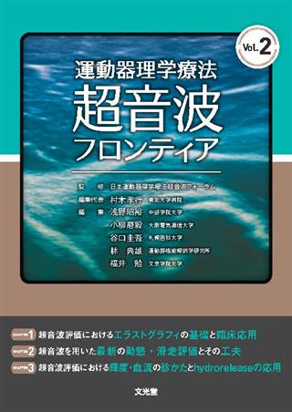
運動器理学療法超音波フロンティア Vol.2

出版社: 文光堂
著者:
発行日: 2022-06-17
分野: 臨床医学:外科  >  リハビリ医学
ISBN: 9784830646010
電子書籍版: 2022-06-17 (第1版第1刷)
書籍・雑誌
≪全国送料無料でお届け≫
取寄せ目安:8~14営業日

4,070 円(税込)

電子書籍
章別単位での購入はできません
ブラウザ、アプリ閲覧

4,070 円(税込)

商品紹介

日本運動器理学療法超音波フォーラムで注目を集めたトピックスを,豊富な図表と動画で解説するシリーズの第2弾.今回は,第2回フォーラムで盛況をみせた「エラストグラフィ」「動態・滑走」「輝度,血流,ハイドロリリース」の3つのシンポジウムに関する17タイトルを収載.超音波画像上で異常があっても,必ずしもそれが痛みの原因とは限らない.本書ではトップランナーの執筆者が,見えているものを理解するために必要な「正しい解釈」を念頭に,最先端の超音波評価を分かりやすく解説した.運動器超音波の「フロンティア」を知りたいセラピスト必携の1冊.

目次

  • CHAPTER 1 超音波評価におけるエラストグラフィの基礎と臨床応用
     Introduction
     1 エラストグラフィの基礎と評価への応用
     2 大腿直筋に加わる力学ストレスに筋内部位差は存在するか?
     3 肩後方タイトネスと肩関節運動時における上腕骨頭の変位方向
     4 上腕三頭筋長頭周辺の組織弾性と肩関節可動域との関係
     5 超音波エラストグラフィによる膝窩筋腱の伸長量評価

    CHAPTER 2 超音波を用いた最新の動態・滑走評価とその工夫
     Introduction
     1 超音波を用いた動態評価の今そしてこれから
     2 超音波診断装置を用いた足部腱滑走距離計測の定量評価法の確立
       ―Thiel法固定遺体,生体を用いた検証―
     3 野球肘に対する肘外反動揺性の定量評価と肘装具の制動効果
       ―超音波診断装置を用いた検証―
     4 超音波診断装置を用いた妊産婦に対する骨盤底筋群評価
     5 ランニング時の大腿部皮膚の挙動について

    CHAPTER 3 超音波評価における輝度・血流の診かたとhydroreleaseの応用
     Introduction
     1 超音波による血流評価の基礎と臨床応用
     2 超音波エコー輝度を用いた筋質評価とその応用
     3 投球障害肩を超音波とhydroreleaseで評価する―可動域制限に着目して―
     4 肩腱板断裂修復術後症例の超音波血流評価

この書籍の参考文献

参考文献のリンクは、リンク先の都合等により正しく表示されない場合がありますので、あらかじめご了承下さい。

本参考文献は電子書籍掲載内容を元にしております。

CHAPTER 1 超音波評価におけるエラストグラフィの基礎と臨床応用

P.12 掲載の参考文献
1) Chino K, et al : Reliability and validity of quantifying absolute muscle hardness using ultrasound elastography. PLoS One 7 : e45764, 2012
2) Shimoyama D, et al : Reliability of shoulder muscle stiffness measurement using strain ultrasound elastography and an acoustic coupler. J Med Ultrason (2001) 48 : 91-96, 2021
3) Muraki T, et al : Ultrasound elastography-based assessment of the elasticity of the supraspinatus muscle and tendon during muscle contraction. J Shoulder Elbow Surg 24 : 120-126, 2015
4) Yanagisawa O, et al : Evaluation of human muscle hardness after dynamic exercise with ultrasound real-time tissue elastography : a feasibility study. Clin Radiol 66 : 815-819, 2011
5) Drakonaki EE, et al : Real-time ultrasound elastography of the normal Achilles tendon : reproducibility and pattern description. Clin Radiol 64 : 1196-1202, 2009
6) Hatta T, et al : In vivo measurement of rotator cufftendon strain with ultrasound elastography : an investigation using a porcine model. J Ultrasound Med 33 : 1641-1646, 2014
7) Ophir J, et al : Elastography : a quantitative method for imaging the elasticity of biological tissues. Ultrason Imaging 13 : 111-134, 1991
8) Ishikawa H, et al : Noninvasive assessment of the activity of the shoulder girdle muscles using ultrasound real-time tissue elastography. J Electromyogr Kinesiol 25 : 723-730, 2015
9) Ishikawa H, et al : Differences in scapular motion and parascapular muscle activities among patients with symptomatic and asymptomatic rotator cufftears, and healthy individuals. JSES Int 5 : 238-246, 2021
10) Ishikawa H, et al : Changes in the shoulder muscle activities and glenohumeral motion after rotator cuffrepair : an assessment using ultrasound real-time tissue elastography. J Shoulder Elbow Surg 30 : 2577-2586, 2021
11) Johansson H, et al : Pathophysiological mechanisms involved in genesis and spread of muscular tension in occupational muscle pain and in chronic musculoskeletal pain syndromes : a hypothesis. Med Hypotheses 35 : 196-203, 1991
12) Ishikawa H, et al : Changes in stiffness of the dorsal scapular muscles before and after computer work : a comparison between individuals with and without neck and shoulder complaints. Eur J Appl Physiol 117 : 179-187, 2017
13) Szeto GP, et al : A comparison of symptomatic and asymptomatic office workers performing monotonous keyboard work--1 : neck and shoulder muscle recruitment patterns. Man Ther 10 : 270-280, 2005
P.21 掲載の参考文献
1) Gajdosik RL : Passive extensibility of skeletal muscle : review of the literature with clinical implications. Clin Biomech 16 : 87-101, 2001
5) Liu J, et al : Non-invasive Quantitative Assessment of Muscle Force Based on Ultrasonic Shear Wave Elastography. Ultrasound Med Biol 45 : 440-451, 2019
6) Iida N, et al : Relationship between shear modulus and passive tension of the posterior shoulder capsule using ultrasound shear wave elastography : A cadaveric study. J Biomech 99 : 109498, 2020
7) 渡邊航平 : 大腿直筋における区画的な神経筋活動. 日本基礎理学療法学雑誌 19 : 2-7, 2016
8) Hasselman CT, et al : An explanation for various rectus femoris strain injuries using previously undescribed muscle architecture. Am J Sports Med 23 : 493-499, 1995
9) Watanabe K, et al : Task-dependent spatial distribution of neural activation pattern in human rectus femoris muscle. J Electromyogr Kinesiol 22 : 251-258, 2012
10) Pasta G, et al : Sonography of the quadriceps muscle : Examination technique, normal anatomy, and traumatic lesions. J Ultrasound 13 : 76-84, 2010
12) Xu J, et al : Stiffness of individual quadriceps muscle assessed using ultrasound shear wave elastography during passive stretching. J Sport Health Sci 7 : 245-249, 2018
13) Le Sant G, et al : Stiffness mapping of lower leg muscles during passive dorsiflexion. J Anat 230 : 639-650, 2017
14) Hug F, et al : Slack length of gastrocnemius medialis and Achilles tendon occurs at different ankle angles. J Biomech 46 : 2534-2538, 2013
15) Meijer HJ, et al : Myofascial force transmission between antagonistic rat lower limb muscles : effects of single muscle or muscle group lengthening. J Electromyogr Kinesiol 17 : 698-707, 2007
P.31 掲載の参考文献
7) Burkhart SS, et al : The disabled throwing shoulder : spectrum of pathology part I : pathoanatomy and biomechanics. Arthroscopy 19 : 404-420, 2003
11) 福吉正樹ほか : 肩甲上腕関節の拘縮からみた肩関節インピンジメント症候群に対する運動療法 : その評価と治療のコツ. 臨床スポーツ医学 30 : 467-472, 2013
12) Mochizuki T, et al : Humeral insertion of the supraspinatus and infraspinatus. New anatomical findings regarding the footprint of the rotator cuff. J Bone Joint Surg 90-Am : 962-969, 2008
14) 加藤敦夫ほか : 小円筋の形態とその支配神経の解剖学的解析. 肩関節 34 : 301-304, 2010
15) Hamada J, et al : Anatomic study and electromyographic analysis of the teres minor. J Shoulder Elbow Surg 26 : 870-877, 2017
16) 福吉正樹ほか : Shear wave elastographyよりみた小円筋上部筋束と下部筋束の機能的特性.日整超会誌 28 : 16-21, 2016
17) Yamamoto N, et al : Contact between the coracoacromial arch and the rotator cufftendons in nonpathologic situations : A cadaveric study. J Shoulder Elbow Surg 19 : 681-687, 2010
P.41 掲載の参考文献
1) 中川宏樹ほか : 大円筋と上腕三頭筋長頭の筋膜連結を考慮した運動療法の試み : 拘縮肩における検討. 日整超会誌 30 : 22-27,2018
2) 二村涼ほか : 小円筋上部筋束ならびに下部筋束の伸張に伴う上腕三頭筋長頭の組織弾性変化について. 整形リハ会誌 21 : 33-37, 2019
4) Handling MA, et al : The origin of the long head of the triceps : a cadaveric study. J Shoulder Elbow Surg 19 : 69-72,2010
5) Standring S : Gray's anatomy : the anatomical basis of clinical practice. 41 st ed., saunders, London, 824-826, 2016
6) 福吉正樹ほか : 大円筋と上腕三頭筋長頭の解剖学的連結に関する機能的意義とは? -shear wave elastographyを用いた検討-. 日整超会誌 29 : 18-24,2017
7) 加藤敦夫ほか : 小円筋の形態とその支配神経の解剖学的解析. 肩関節 34 : 301-304,2010
8) 林典雄ほか : 後方腱板 (棘下筋・小円筋) と肩関節包との結合様式について. 理学療法学 23 : 522-526, 1996
9) 二村涼ほか : 小円筋と上腕三頭筋長頭の解剖学的連結に関する機能的意義について : shear wave elastographyを用いた検討. 日整超会誌 30 : 34-41,2018
10) 二村涼ほか : 投球障害肩における上腕三頭筋長頭周辺の組織弾性ならびに可動域との関係. 日整超会誌 31 : 50-56,2019
11) Lee SY, et al : Correlation of MR arthrographic findings and range of shoulder motions in patients with frozen shoulder. AJR Am J Roentgenol 198 : 173-179, 2012
12) 皆川洋至 : 保存療法 : サイレント・マニピュレーションを中心に-五十肩 (凍結肩). MB Medical Rehabilitation 157 : 85-90, 2013
13) 林典雄ほか : 等尺性収縮を用いた肩関節ROM訓練. 理学療法 17 : 485-489, 1990
14) Wilk KE, et al : Current concepts in the rehabilitation of the overhead throwing athlete. Am J Sports Med 30 : 136-151,2002
15) 福吉正樹ほか : 肩甲上腕関節の拘縮からみた肩関節インピンジメント症候群に対する運動療法 : その評価と運動療法のコツ. 臨スポーツ医 30 : 467-472, 2013
16) 河端将司ほか : 肩肘痛を有するスポーツ選手の末梢神経障害の捉え方. PTジャーナル 54 : 549-558, 2020
17) Woods R, et al : Can Ultrasound Be Used to Improve the Palpation Skills of Physicians in Training? A Prospective Study. PM R 10 : 730-737, 2018
18) 宮武和馬ほか : 超音波ガイド下インターベンションと理学療法の融合. PTジャーナル 54 : 1002-1009, 2020
P.48 掲載の参考文献
1) 中添悠介ほか : 健常膝における跪きから正座までの動態解析. 臨床バイオメカニクス 38 : 345-351, 2017
2) 木村佳記ほか : 術後リハビリテーション. パーフェクト前十字靱帯再建術 (ACL), 金芳堂, 京都, 246-261, 2020
3) 林典雄 : 膝関節伸展機構の機能解剖と膝関節拘縮治療への展開. 愛知県理学療法学会誌 16 : 8-16, 2004
4) 林典雄 : 膝関節疾患における超音波診断装置の臨床応用. 理学療法学 41 : 188-192, 2014
5) Nakagawa S, et al : Tibiofemoral movement 3 full flexion in the living knee studied by MRI. J Bone Joint Surg Br 82 : 1199-1200, 2000
6) 江玉睦明ほか : 膝窩筋機能の肉眼解剖学的検討. スポーツ傷害 18 : 47-49, 2013
7) 国中優治 : 関節病態運動学機能解剖学的に捉えた膝関節の運動学 (解説). 理学療法 24 : 733-743, 2007
8) Maganaris CN : Tensile properties of in vivo human tendinous tissue. J Biomech 35 : 1019-1027, 2002
9) O'Brien TD, et al : Mechanical properties of the patellar tendon in adults and children. J Biomech 43 : 1190-1195, 2010
10) Rosskopf AB, et al : Comparison of shear wave velocity measurements assessed with two different ultrasound systems in an ex-vivo tendon strain phantom. Skeletal Radiol 45 : 1541-1551, 2016
11) 須田康文ほか : 膝関節外側支持機構の機能解剖学的研究 第2報 膝窩筋腱の形態について. 中部整災誌 35 : 1589-1590, 1992
12) Zeng SX, et al : Anatomic study of popliteus complex of the knee in a Chinese population. Anat Sci Int 86 : 213-218, 2011
13) 椎名毅 : エラストグラフィ-static法とdynamic法-. 乳腺甲状腺超音波医学 4 : 11-15, 2015
14) 山越芳樹ほか : せん断波伝搬による組織弾性計測. 超音波医学 42 : 589-597, 2015
15) 山田大智ほか : 生体膝における膝深屈曲が膝窩筋腱の組織弾性変化に及ぼす影響. 日整超会誌 29 : 26-29, 2017
16) 山田大智ほか : 生体膝における膝深屈曲位での下腿回旋が膝窩筋腱の組織弾性に及ぼす影響. 日整超会誌 30 : 140-145, 2018
17) 須田康文ほか : 膝関節外側支持機構の機能解剖学的研究 第4報 後外側支持機構の形態について. 中部整災誌 36 : 1411-1412, 1993

CHAPTER 2 超音波を用いた最新の動態・滑走評価とその工夫

P.51 掲載の参考文献
1) 小柳磨毅ほか : 理学療法におけるポケットエコー (miruco) の活用. 2021 Book超音波診断 映像情報メディカル増刊号 53, 90-94, 2021
P.61 掲載の参考文献
1) Maganaris CN, et al : In vivo human tendon mechanical properties. J Physiol 521 Pt 1 : 307-313, 1999
2) Mohr L, et al : Use of Reflective Tape to Detect Ultrasound Transducer Movement : A Validation Study. Life (Basel) 11 : 104, 2021
3) Kwah LK, et al : Reliability and validity of ultrasound measurements of muscle fascicle length and pennation in humans : a systematic review. J Appl Physiol 114 : 761-769, 2013
4) Li H, et al : Systematic Performance Evaluation of a Cross-Correlation-Based Ultrasound Strain Imaging Method. Ultrasound Med Biol 42, 2436-2456, 2016
5) Frich LH, et al : Musculoskeletal application and validation of speckle-tracking ultrasonography. BMC Musculoskelet Disord 20 : 192, 2019
6) Ishii Y, et al Dynamic ultrasonography of the medial meniscus during walking in knee osteoarthritis. Knee 27 : 1256-1262, 2020
7) Lin S, et al Effect of real-time ultrasound imaging for biofeedback on trunk muscle contraction in healthy subjects : a preliminary study. BMC Musculoskelet Disord 22 : 142, 2021
P.69 掲載の参考文献
1) 牧昌弘ほか : <特集> 足底腱膜炎の診療-足底腱膜炎の診断. Orthopaedics 31 : 9-16, 2018
2) 高橋謙二ほか : アキレス腱症・アキレス腱周囲炎の診断と治療. 関節外科. 36 : 36-43, 2017
3) 服部惣一 : 足部・足関節疾患と外傷に対する保存療法 Evidence-Based Conservative Treatment 足底腱膜炎に対する保存療法. 臨床整形外科 54 : 145-151. 2019
4) 熊井司 : 超音波 (2) : 足関節・足部慢性疾患における超音波画像. MB Orthopaedics 27 : 71-79, 2014
5) 平田正 : 筋・腱損傷と回復過程での超音波利用. MB Med Reha 216 : 71-76, 2017
6) 林典雄 : 理学療法士における超音波画像診断装置の可能性-超音波評価と運動療法とを一直線でつなぐ. 理学療法学 44 : 26-31, 2017
7) Kelly E, et al : Ultrasound assessment of extensor pollicis brevis tendon excursion in different wrist positions in healthy people. J Hand Ther 32 : 375-381, 2018
8) Korstanje JW, et al. : Ultrasonographic assessment of flexor tendon mobilization : effect of different protocols on tendon excursion. J Bone Joint Surg Am 94 : 394-402, 2012
9) 松井智裕ほか : 下腿から足における超音波の利用. MB Med Reha 216 : 16-24, 2017
10) 松井智裕ほか : 足・足関節の画像診療. 関節外科 37 : 60-67, 2018
11) Korstanje JW, et al : Development and validation of ultrasound speckle tracking to quantify tendon displacement. J Biomech 43 : 1373-1379, 2010
12) Lyman J, et al : Strain behavior of the distal achilles tendon : implications for insertional achilles tendinopathy. Am J Sports Med 32 : 457-461, 2004
13) Eisma R, et al : From formalin to Thiel embalming : What changes? One anatomy department's experiences. Clin Anat 26 : 564-571. 2013
14) 谷口達也ほか : 超音波診断装置による足部腱滑走距離の評価-Thiel法固定標本を用いた妥当性・信頼性検証-. 日足外科会誌 40 : 147-150, 2019
15) Landis JR, et al : The measurement of observer agreement for categorical data. Biometrics 33 : 159-174, 1977
16) 谷口達也ほか : 超音波診断装置による足部腱滑走距離計測-異なる足関節肢位の影響-. 日足外科会誌 41 : 118-121, 2020
17) Hintermann B, et al. : Foot movement and tendon excursion : an in vitro study. Foot Ankle Int 15 : 386-395, 1994
P.76 掲載の参考文献
1) 全日本野球協会ほか : 平成28年度中学野球 (軟式・硬式) 実態調査 報告書, 2017
2) Pappas AM : Elbow problems associated with baseball during childhood and adolescence. Clin Orthop Relat Res 164 : 30-41, 1982
3) Fleisig GS, et al : Kinetics of baseball pitching with implications about injury mechanisms. Am J sports Med 23 : 233-239, 1995
4) 彌富雅信ほか : 野球競技者の肘関節内側上顆下端障害と肘関節の形態・機能について. 日本肘関節学会雑誌 22 : 64-67, 2015
5) Ellenbecker TS, et al : Medial elbow joint laxity in professional baseball pitchers. Am J Sports Med 26 : 420-424, 1998
6) Hendawi TK, et al : Medial elbow stability assessment after ultrasound-guided ulnar collateral ligament transection in a cadaveric model : ultrasound versus stress radiography. J Shoulder Elbow Surg 28 : 1154-1158, 2019
7) 大西寛之ほか : 超音波画像を用いた肘外反制動テーピングの効果検証.日本臨床スポーツ医学会誌 25 : S264, 2017
8) Sasaki J, et al : Ultrasonographic assessment of the ulnar collateral ligament and medial elbow laxity in college baseball players. J Bone Joint Surg Am 84 : 525-531, 2002
9) 坂田淳ほか : 内側野球肘患者における超音波検査を用いた肘外反不安定性の経時的変化とX線所見との関連, 日本肘関節学会雑誌 18 : 29-32, 2011
10) Otoshi K, et al : Ultrasonographic assessment of the flexor pronator muscles as a dynamic stabilizer of the elbow against valgus force. Fukushima Jour Med Sci 60 : 123-128, 2014
11) Hattori H, et al : The effects of elbow bracing on medial elbow joint space gapping associated with repetitive throwing in high school baseball players. Orthop J Sports Med 5 : 2325967117702361, 2017
14) Marshall NE, et al : Ulnar collateral ligament and elbow adaptations in high school baseball pitchers. Sports Health 7 : 484-488, 2015
15) 今高康詞ほか : 超音波による側臥位・肩3rd positionでの肘外反動揺性の定量評価-健常肘における相対的・絶対的信頼性-. 保健医療学学会第9回学術集会, 2018
16) 今高康詞ほか : 新しい素材を用いた野球肘予防装具の制動効果. スポーツ傷害 (J.Sports Injury) 24 : 40-42, 2019
17) 多和田兼章ほか : 成長期野球肘内側部障害における超音波所見と投球再開時期の検討. 中部日本整形外科災害外科学会雑誌 54 : 1049-1050, 2011
P.87 掲載の参考文献
1) 布施陽子 : 妊婦への理学療法の効果と臨床 ; 評価編. 臨床助産ケア 9-10 : 79-88, 2017
2) Lien KC, et al : Levator ani muscle stretch induced by simulated vaginal birth. Obstet Gynecol 103 : 31-40, 2004
3) Dietz HP, et al : Levator trauma after vaginal delivery. Obstet Gynecol 106 : 707-712, 2005
4) Krofta L, et al : Pubococcygeus-puborectalis trauma after forceps delivery : evaluation of the levator ani muscle with 3D/4D ultrasound. Int Urogynecol J Pelvic Floor Dysfunct 20 : 1175-1181, 2009
5) Whittaker J : Abdominal Ultrasound Imaging of Pelvic Floor Muscle Function in Individuals with Low Back Pain. J Man Manip Ther 12 : 44-49, 2004
P.94 掲載の参考文献
1) Klous M, et al : Marker-based reconstruction of the kinematics of a chain of segments : a new method that incorporates joint kinematic constraints. J Biomech Eng 132 : 074501, 2010
2) Lu TW, et al : Bone position estimation from skin marker coordinates using global optimization with joint constraints. J Biomech 32 : 129-134, 1999
3) Lucchetti L, et al : Skin movement artefact assessment and compensation in the estimation of knee-joint kinematics. J Biomech 31 : 977-984, 1998
4) Cappello A, et al : Soft tissue artifact compensation in knee kinematics by double anatomical landmark calibration : performance of a novel method during selected motor tasks. IEEE Trans Biomed Eng 52 : 992-998, 2005
5) Andersen MS, et al : Kinematic analysis of over-determinate biomechanical systems. Comput Methods Biomech Biomed Engin 12 : 371-384, 2009
6) Andersen MS, et al : A linear soft tissue artefact model for human movement analysis : proof of concept using in vivo data. Gait Posture 35 : 606-611, 2012
7) Kratzenstein S, et al : Effective marker placement for functional identification of the centre of rotation at the hip. Gait Posture 36 : 482-486, 2012
8) Dumas R, et al : Soft tissue artifact compensation by linear 3D interpolation and approximation methods. J Biomech 42 : 2214-2217, 2009
9) 福井勉 : 皮膚運動学-機能と治療の考え方, 三輪書店, 東京, 2-131, 2010
10) Fukui T, et al : In which direction does skin move during joint movement?. Skin Res Tech 22 : 181-188, 2016
11) Fukui T, et al : Skin Movement Rules Relative to Joint Motions. Clin Res Foot Ankle 5 : 234, 2017
12) 福井勉 : 皮膚テーピング~皮膚運動学の臨床応用~, 運動と医学の出版社, 神奈川, 1-200, 2014

CHAPTER 3 超音波評価における輝度・血流の診かたと hydrorelease の応用

P.110 掲載の参考文献
1) 井上彰ほか : 鎖骨下動脈における血流速度測定の信頼性と第一肋骨切除術前後の血流速度変化における検討. 整スポ会誌 37 : 53-58, 2017
2) Martinoli C, et al : Gain setting in power Doppler. Radiology 202 : 284-285, 1997
3) Corvino A, et al : Utility of a gel stand-offpad in the detection of Doppler signal on focal nodular lesions of the skin. J Ultrasound 23 : 45-53, 2020
4) Forsgren S, et al : Vascular NK-1 receptor occurrence in normal and chronic painful Achilles and patellar tendons : studies on chemically unfixed as well as fixed specimens. Regul Pept 126 : 173-181, 2005
5) Iwamoto W, et al : Transcatheter arterial embolization of abnormal vessels as a treatment for lateral epicondylitis refractory to conservative treatment : a pilot study with a 2-year follow-up. J Shoulder Elbow Surg 26 : 1335-1341, 2017
6) Zeisig E, et al : Extensor origin vascularity related to pain in patients with Tennis elbow. Knee Surg Sports Traumatol Arthrosc 14 : 659-663, 2006
7) Kanbe K, et al : Inducement of mitogen-activated protein kinases in frozen shoulders. J Orthop Sci 14 : 56-61, 2009
8) Okuno Y, et al : Clinical outcomes of transcatheter arterial embolization for adhesive capsulitis resistant to conservative treatment. J Vasc Interv Radiol 28 : 161-167, 2017
9) Inoue A, et al : Evaluation of hypervascularity in synovitis of the shoulder using ultrasound : Comparison of preoperative ultrasound findings and intraoperative arthroscopic findings. JSES international 2022. in press
10) Davis DE, et al : Classifying glenohumeral synovitis : A novel intraoperative scoring system. J Shoulder Elbow Surg 26 : 2047-2053, 2017
11) Kelley MJ, et al : Frozen shoulder : Evidence and a proposed model guiding rehabilitation. J Orthop Sports Phys Ther 39 : 135-148, 2009
12) Naredo E, et al : Assessment of inflammatory activity in rheumatoid arthritis : a comparative study of clinical evaluation with g rey scale and power Doppler ultrasonography. Ann Rheum Dis 64 : 375-381, 2005
13) 岩崎翼ほか : 初期大腿骨顆部特発性骨壊死における超音波検査の有用性.臨整外 53 : 827-830, 2018
14) 金子貴俊ほか : 特発性大腿骨顆部壊死を伴う膝内側部痛に対する超音波検査機器の有用性. 整・災外 64 : 109-111, 2021
15) Jordan RW et al : The importance of early diagnosis in spontaneous osteonecrosis of the knee - A case series with six year follow-up. Knee 23 : 702-707, 2016
16) Al-Rowaih A, et al : Osteonecrosis of the knee. Diagnosis and outcome in 40 patients. Acta Orthop Scand 62 : 19-23, 1991
17) Okuno Y, et al : Midterm Clinical Outcomes and MR Imaging Changes after Transcatheter Arterial Embolization as a Treatment for Mild to Moderate Radiographic Knee Osteoarthritis Resistant to Conservative Treatment. J Vasc Interv Radiol 28 : 995-1002, 2017
18) 小林匠ほか : 軟部組織損傷・障害の病態とリハビリテーションの考え方. 軟部組織損傷・障害の病態とリハビリテーション. 熊井 司ほか, メジカルビュー, 東京, 2-10, 2021
19) Hand C, et al : Long-term outcome of frozen shoulder. J Shoulder Elbow Surg 17 : 231-236, 2008
20) 井上彰ほか : 肩関節におけるエコーを用いた包括的検査-関節リウマチ, リウマチ性多発筋痛症, 肩関節周囲炎における検討-. 第64回 日本リウマチ総会学術集会 (抄録集), 2020.
21) Camellino D, et al : Imaging of polymyalgia rheumatica : indications on its pathogenesis, diagnosis and prognosis. Rheumatology (Oxford) 51 : 77-86, 2012
22) Liu W, et al : Superb Microvascular Imaging Compared with Contrast-Enhanced Ultrasound for Assessing Laser Ablation Treatment of Benign Thyroid Nodules. Biomed Res Int 23 : 1025657, 2018
P.120 掲載の参考文献
1) Goodpaster BH, et al : Subcutaneous abdominal fat and thigh muscle composition predict insulin sensitivity independently of visceral fat. Diabetes 46 : 1579-1585, 1997
2) Ryan AS, et al : Age-related changes in fat deposition in mid-thigh muscle in women : relationships with metabolic cardiovascular disease risk factors. Int J Obes Relat Metab Disord 23 : 126-132, 1999
3) Maeda H, et al : Quadriceps thickness and echo intensity predict gait independence in individuals with severe and mild hemiparetic stroke. Eur Neurol 83 : 167-173, 2020
4) Fukumoto Y, et al : Skeletal muscle quality assessed from echo intensity is associated with muscle strength of middle-aged and elderly persons. Eur J Appl Physiol 112 : 1519-1525, 2012
5) Taniguchi M, et al : Quantity and quality of the lower extremity muscles in women with knee osteoarthritis. Ultrasound Med Biol 41 : 2567-2574, 2015
6) Yoshiko H, et al : Three-dimensional comparison of intramuscular fat content between young and old adults. BMC Med Imaging 17 : 12, 2017
7) Kelley DE, et al : Skeletal muscle density : effects of obesity and non-insulin-dependent diabetes mellitus. Am J Clin Nutr 54 : 509-515, 1991
8) 田村好史ほか : 高齢者における骨格筋の量および質の変化と疾患リスク. 日本サルコペニア・フレイル学会誌 4 : 39-44, 2020
9) 上住円ほか : 骨格筋の線維化-起源となる細胞とその特性. 実験医学 38 : 2101-2107, 2020
10) Akima H, et al : Quadriceps echo intensity can be an index of muscle size regardless of age in 65 or more years old. Exp Gerontol 138 : 111015, 2020
11) Lopez P, et al : Echo intensity independently predicts functionality in sedentary older men. Muscle Nerve 55 : 9-15, 2017
12) Reimers K, et al : Skeletal muscle sonography : a correlative study of echogenicity and morphology. J Ultrasound Med 12 : 73-77, 1993
13) Pillen S, et al : Skeletal muscle ultrasound : correlation between fibrous tissue and echo intensity. Ultrasound Med Biol 35 : 443-446, 2009
14) Akima H, et al : Intramuscular adipose tissue determined by T1 -weighted MRI at 3 T primarily reflects extramyocellular lipids. Magn Reson Imaging 34 : 397-403, 2016
15) Fiji Downloads (Mac版) https://imagei.net/software/fiji
16) Wong V, et al : Exercise induced changes in echo intensity within the muscle : a brief review. J Ultrasound 23 : 457-472, 2020
P.131 掲載の参考文献
1) 宮下浩二 : 運動連鎖から見た投球動作 : 臨床スポーツ医学 29 : 2012, 55-60, 2012
2) 林典雄 : 烏口肩峰靱帯の超音波観察と拘縮の関連. : 運動療法のための運動器超音波機能解剖-拘縮治療との接点, 杉本勝正, 文光堂, 東京, 32-39, 2015
3) 菅谷啓之 : 競技現場のニーズに応じた知識と技術. 臨床スポーツ医学 32 : 114-119, 2015.
4) 山本智章ほか : 野球肘 ; 上腕骨小頭離断性骨軟骨炎の投球動作. 整・災外 58 : 1007-1015, 2015
5) 我妻浩二ほか : 投球障害の肩以外の身体特性-肩甲骨と股関節に着目して. PTジャーナル 54 : 519-525, 2020
6) 岩本航ほか : 肩関節の可動域制限と肩こり. 臨床スポーツ医学 37 : 202-209, 2020
7) 林典雄 : 肩関節周囲組織の動態と理学療法. PTジャーナル 54 : 1010-1015, 2020
8) 福吉正樹ほか : 肩甲上腕関節の拘縮からみた肩関節インピンジメント症候群に対する運動療法. 臨床スポーツ医学 30 : 467-472, 2013
9) 林典雄ほか : 後方腱板 (棘下筋・小円筋) と肩関節包との結合様式について. 理学療法学 23 : 522-527, 1996
10) 二村涼ほか : 小円筋と上腕三頭筋長頭の解剖学的連結に関する機能的意義について-shear wave elastographyを用いた検討-. 日整超会誌 30 : 34-41, 2018
11) 高村隆 : 投球障害の運動療法. 臨床スポーツ医学 32 : 44-51, 2015
12) Matthew B, et al : Prevalence of Scapular Dyskinesis in Overhead and Nonoverhead Athletes : A Systematic Review. Orthop J Sports Med 4 : 2325967115627608, 2016
13) Pluim BM : Scapular dyskinesis : practical applications. Br J Sports Med 47 : 875-876, 2013
14) 遠藤和博ほか : 上肢と肩甲骨運動から見た投球動作の仕組み. 臨床スポーツ医学 29 : 41-45, 2012
15) 宮下浩二ほか : 投球動作の肩最大外旋角度に対する肩甲上腕関節と肩甲胸郭関節および胸椎の貢献度. 体力科学 58, 379-386, 2009
P.141 掲載の参考文献
1) 大泉尚美ほか : 腱板断裂術後後療法. MB Orthopaedics 25 : 59-66, 2012
2) 南川智彦ほか : 腱板小・中断裂の診断と手術・後療法. 関節外科 37 : 728-736, 2018
3) 皆川洋至ほか : スポーツに役立てる超音波画像診断, 皆川洋至, ブックハウス・エイチディ, 東京, 7, 2010
4) 皆川洋至 : 超音波でわかる運動器疾患-診断のテクニック, 鳥羽清治, メジカルビュー社, 東京, 20, 2010
5) 榮崎彰秀ほか : アキレス腱断裂縫合術後症例の超音波画像診断装置による縫合部血流評価から得られた運動療法の検討. 奈良理学療法学 9 : 15-16, 2016
6) 菅谷啓之ほか : 単層固定法における鏡視下腱板修復術の成績-術後1年のMRI所見と手術成績-. 肩関節 27 : 233-236, 2003
7) 熊谷純ほか : 腱板修復機転における肩峰下滑液包の役割-分子生物学的手法を用いて-. 肩関節 18 : 30-35, 1994
8) Smith MM, et al : Modulation of aggrecan and ADAMTS expression in ovine tendinopathy induced by altered strain. Arthritis Rheum 58 : 1055-1066, 2008
9) 熊谷純ほか : 実験的棘上筋腱損傷の治癒過程. 肩関節 19 : 45-49, 1995
10) 榮崎彰秀ほか : 超音波画像診断装置を用いた肩腱板断裂修復術後症例の経時的血流評価. 臨床雑誌整形外科 70 : 1080-1082, 2019

最近チェックした商品履歴

Loading...