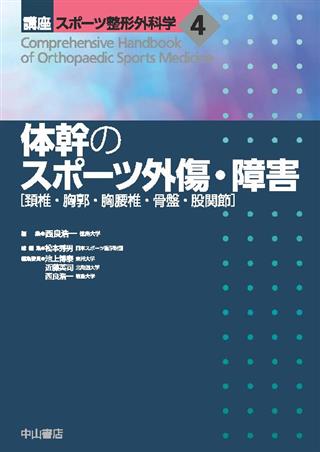
講座 スポーツ整形外科学 4 体幹のスポーツ外傷・障害

出版社: 中山書店
著者:
発行日: 2022-07-30
分野: 臨床医学:外科  >  スポーツ医学
ISBN: 9784521749044
電子書籍版: 2022-07-30 (初版第1刷)
書籍・雑誌
≪全国送料無料でお届け≫
取寄せ目安:8~14営業日

16,500 円(税込)

電子書籍
章別単位での購入はできません
ブラウザ、アプリ閲覧

16,500 円(税込)

商品紹介

スポーツ整形外科領域における頚椎・胸郭・胸腰椎・骨盤・股関節の外傷・障害を取り上げ,それぞれの疾患の特徴や診断の進め方,復帰支援のための診療の実際をまとめた.
スポーツへの復帰に重点をおき,上肢・下肢を含めたさまざまな動作の要である体幹の外傷・障害に対するリハビリテーションや運動療法についても,多くの写真や図を用いて具体的に解説した.
すぐに役立つ必携書.

目次

  • 1章 スポーツによる頚椎の外傷・障害
     頚椎捻挫
     バーナー症候群
     上位頚椎損傷
     中下位頚椎・頚髄損傷
     頚椎椎間板ヘルニアおよび神経根障害
     頚椎症性脊髄症

    2章 スポーツによる胸郭の外傷・障害
     胸骨骨折
     肋骨疲労骨折

    3章 スポーツによる胸腰椎の外傷・障害
     腰部打撲、腰椎捻挫
     成長期腰椎分離症
     成長期骨端輪骨折(小児ヘルニア)
     成人腰椎分離症
     腰椎椎間板ヘルニア
     非特異性腰痛
     椎間板性腰痛
     椎間関節性障害
     仙腸関節障害
     筋性腰痛
     Modic 変化

    4章 スポーツによる骨盤・股関節の外傷・障害
     骨盤裂離骨折
     難治性グロインペイン
     恥骨および坐骨骨折
     寛骨骨折
     FAI(大腿骨寛骨臼インピンジメント)・股関節唇損傷
     弾発股
     梨状筋症候群
     骨盤骨端炎

    5章 体幹運動療法の実際
     Joint by Joint Theory
     Active Corrective Approach
     筋・筋膜スリングとダイアゴナル
     エロンゲーションとアーティキュレーション
     アイソレーションとインテグレーション
     ヨガ―Core Power Yoga CPY
     ピラティス
     モーターコントロールと体幹シナジー

この書籍の参考文献

参考文献のリンクは、リンク先の都合等により正しく表示されない場合がありますので、あらかじめご了承下さい。

本参考文献は電子書籍掲載内容を元にしております。

1章 スポーツによる頚椎の外傷・障害

P.7 掲載の参考文献
1) 米延策雄. スポーツによる頚部の障害・外傷. 二ノ宮節夫ほか (編). 今日の整形外科治療指針. 第5版. 医学書院 ; 2004. p.116-8.
2) 宮崎誠司, 戸松泰介. 柔道. 臨床スポーツ医学 1999 ; 16 : 1275-7.
3) 上田幸夫. レスリング. 臨床スポーツ医学 1999 ; 16 : 1267-70.
4) 藤谷博人ほか. 関東大学アメリカンフットボール秋季公式戦における過去20年間 (1991-2010) の外傷について. 日本臨床スポーツ医学会誌 2012 ; 20 : 550-7.
5) 金岡恒治. 頚椎捻挫. 永廣信治, 西良浩一編. 頭頚部・体幹のスポーツ外傷. 第1版. メジカルビュー ; 2017. p.136-44.
6) 月村泰規. 頚椎捻挫. 中嶋寛之 (監). 新版スポーツ整形外科学. 第1版. 南江堂 ; 2011. p.75-7.
7) 藤谷博人ほか. アメリカンフットボールにおけるネックロールの頚椎制動効果. 整スポ会誌 2001 ; 21 : 5-10.
8) Fujiya H, Goto K. New aspects of microcurrent neuromuscular stimulation in sports medicine. J Phys Fitness Sports Med 2016 ; 5 : 69-72.
9) 藤谷博人. スポーツにおける微弱電流刺激療法. 聖マリアンナ医科大学雑誌 2018 ; 45 : 265-9.
10) Founfelter G. 若年ラグビー選手のための頚椎強化エクササイズ. NSCA JAPAN 2010 ; 17 : 36-41.
11) Grindstaff TL, Potach DH. Prevention of common wresling injuries. Strength Cond 2006 ; 28 : 20-8.
P.13 掲載の参考文献
1) Robertson WC Jr, et al. Upper trunk brachial plexopathy in football players. JAMA 1979 ; 241 : 1480-2.
2) Poindexter DP, et al. Football shoulder and neck injury : A study of the "stinger". Arch Phys Med Rehabil 1984 ; 65 : 601-2.
3) Meyer SA, et al. Cervical spinal stenosis and stingers in collegiate football players. Am J Sports Med 1994 ; 22 : 158-66.
4) 金岡恒治. スポーツにおける頚部のケガ発生メカニズムと予防. Sportsmedicine 2015 ; 27 : 2-10.
5) 阿部均. アメリカンフットボールにおける頚部の外傷・障害 : 頚部のメディカルチェックと医学的な予防対策. 臨床スポーツ医学 1999 ; 16 : 1251-8.
6) Fourre M. On-site management of cervical spine management. Physic Sportsmed 1991 ; 19 : 53-5.
7) Kuhlman GS, et al. The "burner" : A common nerve injury in contact sports. Am Fam Physician 1999 ; 60 : 2035-40, 2042.
8) 仲野春樹ほか. 筋電図検査でC5神経根障害と考えられた遷延性バーナー症候群の1例. 整形外科 2019 ; 70 : 123-5.
9) 真木真一ほか. 大学ラグビー選手を対象にしたバーナー症候群の年間発生調査. 日本臨床スポーツ医会誌 2014 ; 22 : S189.
10) 藤谷博人ほか. 関東大学アメリカンフットボールにおける過去13年間の重症頭部外傷の検討. 整形スポーツ会誌 2007 ; 26 : 257-62.
11) 福井尚志ほか. アメリカンフットボール選手のBurner Syndrome重症例の検討. 日本整形外科スポーツ医学会誌 1993 ; 13 : 17-24.
12) Pavlov H, et al. Cervical spinal stenosis : Determination with vertebral body ratio method. Radiology 1987 ; 164 : 771-5.
13) 阿部均. バーナー症候群. 臨床スポーツ医学 2008 ; 25 : 63-8.
P.21 掲載の参考文献
1) Radcliff KE, et al. In vitro biomechanics of the craniocervical junction : A sequential sectioning of its stabilizing structures. Spine J 2015 ; 15 : 1618-28.
2) Ishii T, et al. Kinematics of the upper cervical spine in rotation : In vivo three-dimensional analysis. Spine (Phila Pa 1976) 2004 ; 29 : E139-44.
3) Miyakoshi N, et al. A nationwide survey on the incidence and characteristics of traumatic spinal cord injury in Japan in 2018. Spinal Cord 2021 ; 59 : 626-34.
4) Miyamoto H, et al. Traumatic anterior atlantoaxial subluxation occurring in a professional rugby athlete : Case report and review of literature related to atlantoaxial injuries in sports activities. Spine (Phila Pa 1976) 2004 ; 29 : E61-4.
5) Werle S, et al. Atlantoaxial instability after a header in an amateur soccer player. Spine (Phila Pa 1976) 2015 ; 40 : E317-20.
6) Rodts GE, et al. Motion-preserving, 2-stage transoral and posterior treatment of an unstable Jefferson fracture in a professional football player. J Neurosurg Spine 2018 ; 28 : 149-53.
7) Alas H, et al. Sports-related cervical spine fracture and spinal cord injury : A review of nationwide pediatric trends. Spine (Phila Pa 1976) 2021 ; 46 : 22-8.
8) Pang D,Wilberger JE Jr. Spinal cord injury without radiographic abnormalities in children. J Neurosurg 1982 ; 57 : 114-29.
9) Pang D. Spinal cord injury without radiographic abnormality in children, 2 decades later. Neurosurgery 2004 ; 55 : 1325-42 ; discussion 1342-3.
10) Trigylidas T, et al. Spinal cord injuries without radiographic abnormality at two pediatric trauma centers in Ontario. Pediatr Neurosurg 2010 ; 46 : 283-9.
11) Mahajan P, et al. Spinal cord injury without radiologic abnormality in children imaged with magnetic resonance imaging. J Trauma Acute Care Surg 2013 ; 75 : 843-7.
12) Banerjee R, et al. Catastrophic cervical spine injuries in the collision sport athlete, part 1 : epidemiology, functional anatomy, and diagnosis. Am J Sports Med 2004 ; 32 : 1077-87.
13) Shimizu T, et al. Scapulohumeral reflex (Shimizu). Its clinical significance and testing maneuver. Spine (Phila Pa 1976) 1993 ; 18 : 2182-90.
14) Spence KF Jr, et al. Bursting atlantal fracture associated with rupture of the transverse ligament. J Bone Joint Surg Am 1970 ; 52 : 543-9.
15) Kandziora F, et al : Spine Section of the German Society for Orthopaedics and Trauma. Treatment of atlas fractures : Recommendations of the Spine Section of the German Society for Orthopaedics and Trauma (DGOU). Global Spine J 2018 ; 8 (2 Suppl) : 5S-11S.
16) Alves OL, et al. Upper cervical spine trauma : WFNS Spine Committee Recommendations. Neurospine 2020 ; 17 : 723-36.
17) Piple A, et al. Cervical spine CT can miss fractures in American football players when protective equipment is in place : Acadaver study. Clin Orthop Relat Res 2021 ; 479 : 2072-80.
18) Torg JS, Ramsey-Emrhein JA. Management guidelines for participation in collision activities with congenital, developmental, or postinjury lesions involving the cervical spine. Clin J Sport Med 1997 ; 7 : 273-91.
19) France JC, et al. Return to play after cervical spine injuries : A consensus of opinion. Global Spine J 2016 ; 6 : 792-7.
20) Nagoshi N, et al. Return to play in athletes with spinal cord concussion : A systematic literature review. Spine J 2017 ; 17 : 291-302.
21) Schroeder GD, et al. Updated return-to-play recommendations for collision athletes after cervical spine injury : A modified delphi consensus study with the Cervical Spine Research Society. Neurosurgery 2020 ; 87 : 647-54.
22) Eddy D, et al. A review of spine injuries and return to play. Clin J Sport Med 2005 ; 15 : 453-8.
23) Wang D, et al. Athletes with musculoskeletal injuries identified at the NFL Scouting Combine and Prediction of Outcomes in the NFL : A systematic review. Orthop J Sports Med 2018 ; 6 : 2325967118813083.
P.41 掲載の参考文献
1) Allen BL, et al. A mechanistic classification of closed, indirect fractures and dislocations of the lower cervical spine. Spine 1982 ; 7 : 1-27.
2) 前田健. 頚椎骨折. 富士川恭輔, 鳥巣岳彦編. 骨折・脱臼. 改訂4版. 南山堂 ; 2018. p.769-818.
3) ASIA and ISCoS International Standards Committee. The 2019 revision of the International Standards for Neurological Classification of Spinal Cord Injury (ISNCSCI) -What's new? Spinal Cord 2019 ; 57 : 815-7.
4) Torg JS, et al. Neurapraxia of the cervical spinal cord with transient quadriplegia. J Bone Joint Surg Am 1986 ; 68 : 1354-70.
5) Maeda T, et al. Soft tissue damage and segmental instability in adult patients with cervical spinal cord injurywithout major bone injury. Spine 2012 ; 37 : E1560-6.
6) 佐々木貴之. 頚髄損傷者の呼吸理学療法. 理学療法 2018 ; 35 : 47-55.
7) 中村濃. 頚髄損傷の動作障害に対する理学療法アプローチ. 理学療法 2010 ; 27 : 118-29.
8) 村井聖ほか. 受傷時年齢が頚髄損傷者のADL獲得に及ぼす影響 : 運動完全麻痺者の残存機能別ADL自立度について. 理学療法学 2020 ; 47S1 : F-47_2.
9) Torg JS, Ramsey-Emrhein JA. Suggested management guidelines for participation in collision activities with congenital, developmental, or postinjury lesions involving the cervical spine. Med Sci Sports Exerc 1997 ; 297 (Suppl) : S256-72.
10) Vaccaro AR, et al. Return to play criteria for the athlete with cervical spine injuries resulting in stinger and transient quadriplegia/paresis. Spine J 2002 ; 2 : 351-6.
11) Cantu RV, Cantu RC. Current thinking : Return to play and transient quadriplegia. Curr Sports Med Rep 2005 ; 41 : 27-32.
12) Torg JS, et al. Injuries to the cervical spine in American football players. J Bone Joint Surg Am 2002 ; 84 : 112-22.
P.54 掲載の参考文献
1) Tong HC, et al. The Spurling test and cervical radiculopathy. Spine 2002 ; 27 : 156-9.
2) Torg JS, et al. Neurapraxia of the cervical spinal cord with transient quadriplegia. J Bone Joint Surg Am 1986 ; 68 : 1354-70.
3) Andrews J, et al. Is return to professional rugby union likely after anterior cervical spinal surgery? J Bone Joint Surg (Br) 2008 ; 90-B : 619-21.
4) Hsu WK. Outcomes following nonoperative and operative treatment for cervical disc herniations in national football league athletes. Spine 2011 ; 36 : 800-5.
5) Adamson TE. Posterior cervical endoscopic laminoforaminotomy for the treatment of radiculopathy in the athlete. Oper Tech Sports Med 2005 ; 13 : 96-100.
6) 中川幸洋, 吉田宗人. アスリートに対する頚椎内視鏡手術. 関節外科 2016 ; 35 : 523-33.
7) 中川幸洋. アスリートに対するMEDシステムを用いた頚椎内視鏡手術. 脊椎脊髄ジャーナル 2018 ; 31 : 209-17.
8) Torg JS, Ramsey-Emrhein JA. Management guidelines for participation in collision activities with congenital, developmental, or postinjury lesions involving the cervical spine. Clin J Sports Med 1997 ; 7 : 273-91.
P.60 掲載の参考文献
1) Berge J, et al. Age-related changes in the cervical spines of front-line rugby players. Am J Sports Med 1999 ; 27 : 422-9.
2) Spurling RG, et al. Lateral rupture of the cervical intervertebral discs : A common cause of shoulder and arm pain. Surg Gynecol Obstet 1944 ; 78 : 350-8.
3) Pavlov H, et al. Cervical spinal stenosis : Determination with vertebral body ratio method. Radiology 1987 ; 164 : 771-5.
4) 阿部均ほか. アメリカンフットボール, ラグビー選手における一過性四肢麻痺の治療経験. 日本整形外科スポーツ医学会雑誌 1998 ; 18 : 71-7.
5) 開地逸朗. 小児の頚椎椎管前後径のX線的観察. 日本外科宝函 1980 ; 49 : 436-53.

2章 スポーツによる胸郭の外傷・障害

P.65 掲載の参考文献
1) 三原和夫. 肋骨・胸骨の骨折. 整形外科MOOK 1980 ; 16 : 191-7.
2) Peek GJ, et al. Isolated sternal fracture : An audit 10 years? experience. Injury 1995 ; 26 : 385-8.
3) Standring S, ed. Gray's Anatomy : The anatomical basis of clinical practice. Churchill Livingstone/Elsevier ; 2008.
4) Watkins R 4th, et al. Stability provided by the sternum and rib cage in the thoracic spine. Spine 2005 ; 30 : 1283-6.
5) Fowler AW. Flexion-compression injury of the sternum. J Bone Joint Surg 1957 ; 39-B : 487-97.
6) Johnson I, Branfoot T. Sternal fracture : A modern review. Arch Emerg Med 1992 ; 10 : 24-8.
7) De Palma AF. The Management of Fractures and Dislocations. Saunders ; 1965.
8) 浅井康文ほか. 外傷性胸骨骨折に対する観血的胸骨固定術. 日本外傷研究会誌 1990 ; 3 : 257-62.
9) 森川洋匡ほか. 胸骨骨折に対する観血的固定術を施行した症例の検討. 日本呼吸器外科学会雑誌 2004 ; 18 : 816-20.
10) 簗瀬誠ほか. 胸骨骨折高度転位例の治療経験. 日職災医誌 2002 ; 50 : 373-7.
P.71 掲載の参考文献
1) 有馬亨. 疲労骨折. 整形外科MOOK 1983 ; 27 : 129-40.
2) 杉浦保夫ほか. スポーツ選手に認められた疲労骨折 (過労性骨障害). 災害医学 1977 ; 20 : 939-49.
3) 藤原明ほか. 野球によって生じた肋骨疲労骨折の3症例. 整形外科と災害外科 1996 ; 45 : 478-81.
4) 佐藤士郎ほか. 肋骨疲労骨折の発生メカニズムについて. 日本整形外科学会雑誌 1991 ; 9 : 708-18.
5) Lee AD, et al. Golf-related stress fractures : A structured review of the literature. J Can Chiropr Assoc 2009 ; 53 : 290-9.
6) Satou S, et al. The mechanism of fatigue fracture of the ribs. J Jpn Orthop Assoc 1991 ; 65 : 708-19.
7) 田中直史ほか. ゴルフスイングにおける肩甲胸郭関節の働きについて. 整形外科スポーツ医学会誌 1994 ; 14 : 79-88.
8) 井上智雄ほか. 野球選手の利き腕側に発生した肋骨疲労骨折の2症例. 臨床整形外科 1986 ; 21 : 1179-81.
9) 柚木修ほか. アンダースロー投手の利き腕側肋骨疲労骨折 (原因と予防). 整形外科スポーツ医学会誌 1986 ; 5 : 27-31.
10) Aitken AP, et al. Fracture of the first rib due to muscle pull. N Engl J Med 1939 ; 220 : 1063-4.
11) O'neal M, et al. First rib stress fracture and pseudoarthrosis in the adolescent athlete : The role of costosternal anatomy. Clin J Sport Med 2009 ; 19 : 65-7.
12) 上野尚ほか. ソフトボールによる第1肋骨疲労骨折の1症例. 整形外科スポーツ医学会誌 1987 ; 6 : 81-4.
13) 内田繕博. スポーツ選手に発症した第1肋骨疲労骨折の2例. JOSKAS 2013 ; 38 : 789-93.
14) 金村斉ほか. 弓道選手に生じた第1肋骨疲労骨折の1例. 整形外科 2013 ; 64 : 974-7.
15) 五嶋謙一ほか. 第1肋骨疲労骨折の臨床的特徴. 日本臨床スポーツ医学会誌 2016 ; 24 : 24-8.
16) 皆川洋至ほか. 肋骨骨折診断における単純X線検査と超音波検査の比較. 日本整形外科超音波研究会誌 2009 ; 21 : 46-50.
17) Mithofer K, et al. Pseudarthrosis of the first rib in the overhead athlete. Br J Sports Med 2004 ; 38 : 221-2.
18) Terabayashi N, et al. Nonunion of a first rib fracture causing thoracic outlet syndrome in a basketball player : A case report. J Shoulder Elbow Surg 2010 ; 19 : 20-3.

3章 スポーツによる胸腰椎の外傷・障害

P.79 掲載の参考文献
1) TOKYO2020オリンピックパラリンピックホームページ. https://olympics.com/ja/olympic-games/tokyo-2020
2) Anderson JE ed. 森田茂, 楠豊訳. Grant's ATLAS OF ANATOMY グラント解剖学図譜. 第3版. 医学書院 ; 1996.
3) 日本整形外科学会, 日本腰痛学会監修. 腰痛診療ガイドライン 2019. 改訂第2版. 南江堂 ; 2019.
4) Chou R, et al. Diagnosis and treatment of low back pain : A joint clinical practice guideline from the American College of Physicians and the American Pain Society. Ann Internal Med 2007 ; 147 : 478.
5) 佐々尾宙ほか. 青年期における腰椎外傷・障害の疫学的研究. 臨床スポーツ医学 2012 ; 29 : 767-74.
6) 日本外傷学会, 日本救急医学会監修. 外傷初期診療ガイドラインJATEC. 改訂第6版. へるす出版 ; 2021.
P.86 掲載の参考文献
1) Sakai T, et al. Incidence of lumbar spondylolysis in the general population in Japan based on multidetector computed tomography scans from two thousand subjects. Spine (Phila Pa 1976) 2009 ; 34 : 2346-50.
2) Aoki Y, et al. Prevalence of lumbar spondylolysis and spondylolisthesis in patients with degenerative spinal disease. Sci Rep 2020 ; 10 : 6739.
3) 青木保親. 診断の最前線-痛い分離と痛くない分離. 整形・災害外科 2020 ; 63 : 1475-9.
4) 酒井紀典, 西良浩一. 小児期腰椎分離症. MB Orthop 2015 ; 28 : 120-6.
5) Sugiura S, et al. Characteristics of low back pain in adolescent patients with early-stage spondylolysis evaluated using a detailed visual analogue scale. Spine (Phila Pa 1976) 2015 ; 40 : E29-34.
6) Yamane T, et al. Early diagnosis of lumbar spondylolysis by MRI. J Bone Joint Surg Br 1993 ; 75 : 764-8.
7) Sairyo K, et al. MRI signal changes of the pedicle as an indicator for early diagnosis of spondylolysis in children and adolescents : A clinical and biomechanical study. Spine (Phila Pa 1976) 2006 ; 31 : 206-11.
8) Sairyo K, et al. Conservative treatment for pediatric lumbar spondylolysis to achieve bone healing using a hard brace : What type and how long? : Clinical article. J Neurosurg Spine 2012 ; 16 : 610-4.
9) Dai LY. Disc degeneration in patients with lumbar spondylolysis. J Spinal Disord 2000 ; 13 : 478-86.
10) 畠山健次. 運動療法のピットフォール発育期腰椎分離症. 臨床スポーツ医学 2020 ; 37 : 1060-5.
11) 青木保親ほか. 腰椎分離症のサイエンス : フィールド復帰のための方程式中・高校生の復帰方程式. 臨床スポーツ医学 2019 ; 36 : 1172-7.
13) 石濱嘉絋ほか. 腰椎分離症に対するSmiley Face Rod法-手術治療の変遷と低侵襲化. 臨床スポーツ医学 2019 ; 36 : 1134-9.
14) 杉浦史郎ほか. 発育期腰椎分離症-装具療法中のエクササイズ. 臨床スポーツ医学 2016 ; 33 : 994-8.
15) Sakai T, et al. Conservative treatment for bony healing in pediatric lumbar spondylolysis. Spine (Phila Pa 1976) 2017 ; 42 : E716-20.
P.96 掲載の参考文献
1) Sairyo K, et al. Three dimensional finite element analysis of the pediatric lumber spine. Part I : Pathomechanism of apophyseal bony ring fracture. Eur Spine J 2006 ; 15 : 923-9.
2) Sairyo K, et al. The pathomechanism of isthmic lumbar spondylolisthesis. A biomechanical study in immature calf spines. Spine 1998 ; 23 : 1442-6.
3) Rajasekaran S, et al. ISSLS prize winner : A study of diffusion in human lumbar discs : A serial magnetic resonance imaging study documenting the influence of the endplate on diffusion in normal and degenerate discs. Spine 2004 ; 29 : 2654-67.
4) Bick EM, Copel JW. The ring apophysis of the human vertebra ; Contribution to human osteogeny. II. J Bone Joint Surg Am 1951 ; 33-A : 783-7.
5) Moore RJ, et al. The vertebral endplate, disc degeneration, disc regeneration. Eur Spine J 2006 ; 15 : S333-7.
6) Rajasekaran S, et al. The anatomy of failure in lumbar disc herniation : An in vivo, multimodal, prospective study of 181 subjects. Spine 2013 ; 38 : 1491-500.
7) Cook G. PART I スポーツの動作. 身体のシステム. 石塚利光ほか監修. アスレティックボディインバランス─パフォーマンスに最も適した動作スキルとコンディショニング. ブックハウス・エイチディ ; 2011. p.14-5.
8) Cook G. 関節別アプローチの概念. 中丸宏二ほか監訳. ムーブメント─ファンクショナルムーブメントシステム. ナップ ; 2014. p.308-10.
9) Tamaki S, et al. Lumbar posterior apophyseal ring fracture combined with spondylolysis in pediatric athletes : Case series of three patients. JBJS case connect 2016 ; 6 : e64.
P.103 掲載の参考文献
1) Sakai T, et al. Incidence of lumbar spondylolysis in the general population in Japan based on multidetector computed tomography scans from two thousand subjects. Spine (Phila Pa 1976) 2009 ; 34 : 2346-50.
2) Tezuka F, et al. Etiology of adult-onset stress fracture in the lumbar spine. Clin Spine Surg 2017 ; 30 : E233-8.
3) Sairyo K, et al. Spondylolysis fracture angle in children and adolescents on CT indicates the facture producing force vector : Abiomechanical rationale. Internet J Spine Surg 2004 ; 1.
4) Terai T, et al. Spondylolysis originates in the ventral aspect of the pars interarticularis : A clinical and biomechanical study. J Bone Joint Surg Br 2010 ; 92 : 1123-7.
5) Sairyo K, et al. Painful lumbar spondylolysis among pediatric sports players : A pilot MRI study. Arch Orthop Trauma Surg 2011 ; 131 : 1485-9.
6) Hirano A, et al. Charactaristics of clinical and imaging findings in adlescent lumbar spondylolysis associated with sports activities. J Spine 2012 ; 1 : 5.
7) Sairyo K, et al. Conservative treatment for pediatric lumbar spondylolysis to achieve bone healing using a hard brace : What type and how long? : Clinical article. J Neurosurg Spine 2012 ; 16 : 610-4.
8) Sakai T, et al. Conservative treatment for bony healing in pediatric lumbar spondylolysis. Spine (Phila Pa 1976) 2017 ; 42 : E716-20.
9) Konz RJ, et al. The pathomechanism of spondylolytic spondylolisthesis in immature primate lumbar spines in vitro and finite element assessments. Spine 2001 ; 26 : E38-49.
10) Morscher E, et al. Surgical treatment of spondylolisthesis by bone grafting and direct stabilization of spondylolysis by means of a hook screw. Arch Orthop Trauma Surg 1984 ; 103 : 175-8.
11) Salib RM, Pettine KA. Modified repair of a defect in spondylolysis or minimal spondylolisthesis by pedicle screw, segmental wire fixation, and bone grafting. Spine 1993 ; 18 : 440-3.
13) Songer MN, Rovin R. Repair of the pars interarticularis defect with a cable-screw construct. A preliminary report. Spine (Phila Pa 1976) 1998 ; 23 : 263-9.
14) Gillet P,P etit M. Direct repair of spondylolysis without spondylolisthesis, using a rod-screw construct and bone grafting of the pars defect. Spine (Phila Pa 1976) 1999 ; 24 : 1252-6.
15) Ulibarri JA, et al. Biomechanical and clinical evaluation of a novel technique for surgical repair of spondylolysis in adolescents. Spine (Phila Pa 1976) 2006 ; 31 : 2067-72.
16) Sairyo K, et al. A new endoscopic technique to decompress lumbar nerve roots affected by spondylolysis. J Neurosurg 2003 ; 98 : 290.
P.113 掲載の参考文献
1) Olmarker K, et al. Tumor necrosis factor alpha and nucleus pulposus-induced nerve root injury. Spine 1998 ; 23 : 2538-44.
2) Macnab I. Bachache. 2nd ed. Williams & Wilkins ; 1990.
3) Gay BL, et al. Disc herniation in the National Football League. Spine (Phila Pa 1976) 2013 ; 38 : 1934-8.
4) Hangai M, et al. Lumbar intervertebral disk degeneration in asthletes. Am J Sports Med 2009 ; 37 : 149-55.
5) Huang W, et al. Is smoking a risk factor for lumbar disc herniation? Eur Spine J 2016 ; 25 : 168-76.
6) Schumann B, et al. Lifestyle factors and lumbar disc disease : Results of a German multi-center casecontrol study (EPILIFT). Arthritis Res Ther 2010 ; 12 : R193.
7) Nachemson AL. Disc pressure measurements. Spine 1981 ; 6 : 93-7.
8) Barker PJ, et al. Effects of tensioning the lumbar fasciae on segmental stiffness during flexion and extension : Young investigator award winner. Spine 2006 ; 31 : 397-405.
9) Cook G 著, 中丸宏二ほか監訳. 付録1. 関節別アプローチの概念, 付録2. 関節別アプローチの詳細. ムーブメント─ファンクショナルムーブメントシステム : 動作のスクリーニング, アセスメント, 修正ストラテジー. ナップ ; 2014. p.308-17.
10) Haro H. Translational research of herniated discs : Current status of diagnosis and treatment. J Orthop Sci 2014 ; 19 : 515-20.
11) Sairyo K, et al. State-of-the-art transforaminal percutaneous endoscopic lumbar surgery under local anesthesia : Discectomy, foraminoplasty, and ventral facetectomy. J Orthop Sci 2018 ; 23 : 229-36.
12) Reiman MP, et al. Return to play after open and microdiscectomy surgery versus conservation treatment for lumbar disc herniation : A systematic reviewwith meta-analysis. Br J Sports Med 2016 ; 50 : 221-30.
13) Cook RW, et al. Return to sport after lumbar spine surgery. Clin Sport Med 2016 ; 35 : 609-19.
14) Yamaya S, et al. Early outcomes of transforaminal percutaneous endoscopic lumbar discectomy for high school athletes with herniated nucleus pulposus of the lumbar spine. J Pediatr Orthop B 2020 ; 29 : 599-606.
P.120 掲載の参考文献
1) Deyo RA, et al. What can the history and physical examination tell us about low back pain? JAMA 1992 ; 268 : 760-5.
2) Suzuki H, et al. Diagnosis and characters of non-specific low back pain in Japan : The Yamaguchi low back pain study. PLoS One 2016 ; 11 : e0160454.
3) 日本整形外科学会, 日本腰痛学会監. 腰痛診療ガイドライン 2012. 南江堂 ; 2012.
4) Mera Y, et al. Association between types of Modic changes in the lumbar region and lowback pain in a large cohort : The Wakayama spine study. Eur Spine J 2021 ; 30 : 1011-7.
5) Takenaka S, et al. Neurological manifestations of thoracic myelopathy. Arch Orthop Trauma Surg 2014 ; 134 : 903-12.
6) Kato K, et al. Ossification of the ligamentum flavum in the thoracic spine mimicking sciatica in a young baseball pitcher : A case report. Fukushima J Med Sci 2021 ; 67 : 33-7.
7) Polgar F. Uber interakuelle wirbelverkalkung. Fortschr Geb Rongenstr Nuklearmed Erganzungsband 1920 ; 40 : 292-8.
8) 安斉直. 肥厚硬化セル黄靱帯ニ依ル脊髄神経根圧迫症例. 日本整形外科学会雑誌 1938 ; 13 : 305-16.
9) 山口博ほか. 脊髄腫瘍症状を呈した黄色靱帯骨化の1例. 整形外科 1960 ; 11 : 951-7.
10) Mori K, et al. Prevalence, distribution, and morphology of thoracic ossification of the yellow ligament in Japanese : Results of CT-based cross-sectional study. Spine (Phila Pa 1976) 2013 ; 38 : E1216-22.
11) Liang H, et al. Epidemiology of ossification of the spinal ligaments and associated factors in the Chinese population : A cross-sectional study of 2000 consecutive individuals. BMC Musculoskelet Disord 2019 ; 20 : 253.
12) Kim SI, et al. Prevalence and related clinical factors of thoracic ossification of the ligamentum flavum : A computed tomography-based cross-sectional study. Spine J 2018 ; 18 : 551-7.
13) Adams MA, Hutton WC. The effect of posture on the role of the apophysial joints in resisting intervertebral compressive forces. J Bone Joint Surg Br 1980 ; 62 : 358-62.
14) Morimoto M, et al. Age-related changes in axial and sagittal orientation of the facet joints : Comparison with changes in degenerative spondylolisthesis. J Orthop Sci 2019 ; 24 : 50-6.
P.127 掲載の参考文献
1) 半谷美夏. トップアスリートにおける腰痛診療. 山下敏彦ほか編. プロフェッショナル腰痛診療. 中外医学社 ; 2018. p.148-61.
2) 松野丈夫, 中村利孝総編集. 標準整形外科学. 第12版. 医学書院 ; 2014.
3) 青木保親ほか. 椎間板性腰痛. 西良浩一編. 極めるアスリートの腰痛-100%を超える復帰. 文光堂 ; 2018. p.73-91.
4) Nachemson AL. Disc pressure measurements. Spine 1981 ; 6 : 93-7.
5) April C, et al. High-intensity zone : A diagnostic sign of painful lumbar disc on magnetic resonance imaging. Br J Radiol 1992 ; 65 : 361-9.
6) Shellhas KP, et al. Lumbar disc high-intensity zone. Correlation of magnetic resonance imaging and discography. Spine 1996 ; 21 : 79-86.
7) Saifuddin A, et al. The value of lumbar spine magnetic resonance imaging in the demonstration of annular tear. Spine 1998 ; 23 : 453-7.
8) Wang ZX, et al. High intensity zone (HIZ) of lumbar intervertebral disc on T1-weighted magnetic resonance images : Spatial distribution, and correlation of distribution with low back pain (LBP). Eur Spine J 2012 ; 21 : 1311-5.
9) Carragee EJ, et al. Lumbar high-intensity zone and discography in subjects without low back pain problems. Spine 2000 ; 25 : 2987-92.
10) Liu C, et al. Quantitative estimation of the high-intensity zone in the lumbar spine : Comparison between the symptomatic and asymptomatic population. Spine J 2014 ; 14 : 391-6.
11) Jha SC, et al. Clinical significance of high-intensity zone for discogenic low back pain : A review. J Med Invest 2015 ; 63 : 1-7.
12) Vanharanta H, et al. The relationship of provocation to lumbar disc deterioration a seen by CT/discography. Spine 1987 ; 12 : 295-8.
13) Vanharanta H, et al. Disc deterioration in low back syndrome. A prospective, multi-center CT/discography study. Spine 1998 ; 13 : 1349-51.
14) Cook G, et al. Understanding corrective strategies. Movement. On Target Publications ; 2010. p.217-32.
15) Cook G, et al. The joint-by joint concept. Movement. On Target Publications ; 2010. p.319-21.
16) Tsou PM, et al. Posterolateral transforaminal selective endoscopic discectomy and thermal annuloplasty for clinical lumbar discogenic pain : A minimal access visualized intradiscal surgical procedure. Spine J 2004 ; 4 : 564-73.
17) Manabe H, et al. Thermal annuloplasty using percutaneous endoscopic discectomy for elite athletes with discogenic low back pain. Neurol Med Chir 2019 ; 59 : 48-53.
P.136 掲載の参考文献
1) Panjabi MM, et al. Articular facets of the human spine. Quantitative three-dimensional anatomy. Spine (Phila Pa 1976) 1993 ; 18 : 1298-310.
2) Manchikanti L, et al. Prevalence of lumbar facet joint pain in chronic low back pain. Pain Physician 1999 ; 2 : 59-64.
3) Schwarzer AC, et al. Clinical features of patients with pain stemming from the lumbar zygapophysial joints. Is the lumbar facet syndrome a clinical entity? Spine (Phila Pa 1976) 1994 ; 19 : 1132-7.
4) Adams MA, Hutton WC. The effect of posture on the role of the apophysial joints in resisting intervertebral compressive forces. J Bone Joint Surg [Br] 1980 ; 62 : 358-62.
5) Sairyo K, et al. Spondylolysis fracture angle in children and adolescents on CT indicates the facture producing force vector : A biomechanical rationale. Internet J Spine Surg 2005 ; 1.
7) 眞鍋裕昭, 西良浩一. 部位ごとのスポーツ外傷・障害脊椎のスポーツ障害. 関節外科基礎と臨床 2019 ; 38 : 1181-9.
8) Bogduk N. Nerves of the lumber spine. Clinical Anatomy of the Lumbar Spine and Sacrum. 3rd edition. Churchill Livingstone ; 1997. p.127-44.
9) Kamada M, et al. Full-endoscopic decompression of foraminal stenosis caused by facet hypertrophy contralateral to the dominant hand in a baseball pitcher : A case report. NMC Case Report Journal 2020 ; 7 : 173-7.
10) Cook G. ムーブメント─ファンクショナルムーブメントシステム : 動作スクリーニング, アセスメント, 修正ストラテジー. ナップ ; 2014.
11) Bockenhauer SE, et al. Measuring thoracic excursion : Reliability of cloth tape measure technique. J Am Osteopath Assoc 2007 ; 105 : 191-6.
12) Kendall FP, et al. Muscles Testing and Function. 5th edition. Williams & Wilkins ; 2005.
13) Clapis PA, et al. Reliability of inclinometer and goniometric measurements of hip extension flexibility using the modified Thomas test. Physiother Theory Pract 2008 ; 24 : 135-41.
14) Peeler J, et al. Using digital photography to document rectus femoris flexibility : A reliability study of the modified Thomas test. Physiother Theory Pract 2013 ; 29 : 319-27.
15) White AA, et al. Clinical Biomechanics of the Spine. 2nd edition. Lippincott Williams & Wilkins ; 1990.
16) Bergmark A. Stability of the lumbar spine. A study in mechanical engineering. Acta Orthop Scand Suppl 1989 ; 230 : 1-54.
17) Luomajoki H, et al. Reliability of movement control test in the lumbar spine. BMC Musculoskelet Disord 2007 ; 8 : 90.
18) Kendall FP, et al. 筋・機能とテスト-姿勢と痛み. 第4版. 西村書店 ; 2006.
19) Schwarzer AC, et al. The false-positive rate of uncontrolled diagnostic blocks of the lumbar zygapophysial joints. Pain 1994 ; 58 : 195-200.
20) van Kleef M, et al. Randomized trial of radiofrequency lumbar facet denervation for chronic low back pain. Spine 1999 ; 24 : 1937-42.
21) Leclaire R, et al. Radiofrequency facet joint denervation in the treatment of low back pain : A placebo-controlled clinical trial to assess efficacy. Spine 2001 ; 26 : 1411-7.
22) 藤原務. スポーツ. 柿崎藤泰編. 胸郭運動システムの再建法. 第2版. ヒューマン・プレス ; 2017. p.210-6.
23) Aasa B, et al. Individualized low-load motor control exercises and education versus a high-load lifting exercise and education to improve activity, pain intensity, and physical performance in patients with low back pain : A randomized controlled trial. J Orthop Sports Phys Ther 2015 ; 45 : 77-85.
P.145 掲載の参考文献
1) Murakami E. Sacroiliac joint disorder. Accurately Diagnosing Low Back Pain. Springer ; 2018. p.7-10.
2) Bernard TN, et al. The sacroiliac joint syndrome. Pathophysiology, diagnosis and management. Frymoyer JW ed. The Adult Spine : Principles and Practice. Lippincott-Raven Publishers ; 1997. p. 2343-63.
3) Toyohara R, et al. Finite element analysis of load transition on sacroiliac joint during bipedal walking. Sci Rep 2020 ; 10 : 13683.
4) Bernard TN, et al. Recognizing specific characteristics of nonspecific low back pain. Clin Orthop Relate Res 1985 ; 217 : 266-80.
5) 成田崇矢ほか. 徒手療法を用いた腰痛の病態評価の試み. 日本臨床スポーツ医学会誌 2017 ; 37 : 22-6.
6) 黒澤大輔, 村上栄一. 仙腸関節痛の発生・慢性化のメカニズム. J Spine Res 2021 ; 12 : 808-13.
7) Murakami E, et al. Treatment strategy for sacroiliac joint-related pain at the posterior superior iliac spine. Clin Neurol Neurosurg 2018 ; 165 : 43-6.
8) 博田節夫編著. 関節運動学的アプローチ博田法. 第2版. 医歯薬出版 ; 2007. p.160.
9) 黒澤大輔, 村上栄一. 腰痛に対する徒手検査. 関節外科 2020 ; 39 : 6-16.
10) 黒澤大輔, 村上栄一. 仙腸関節ブロック. 徳橋泰明編. 整形外科医のための局所麻酔法・ブロック療法. 第2版. メジカルビュー社 ; 2019. p.178-95.
11) Lee JJ, et al. Pain relief scale is more highly correlated with numerical rating scale than with visual analogue scale in chronic pain patients. Pain Physician 2015 ; 18 : E195-200.
12) Tofuku K, et al. The diagnostic value of single-photon emission computed tomography/computed tomography for severe sacroiliac joint dysfunction. Eur Spine J 2015 ; 24 : 859-63.
13) 黒澤大輔ほか. 仙腸関節障害に対する骨SPECT/CT検査の有用性. 整形外科 2021 ; 72 : 249-53.
14) Cusi M, et al. Metabolic disturbances identified by SPECT-CT in patients with a clinical diagnosis of sacroiliac joint incompetence. Eur Spine J 2013 ; 22 : 1674-82.
15) 金岡恒治. アスリートの腰痛に対する評価・診断. 村上栄一編. 長引く腰痛はこうして治せ. 医事新報社 ; 2020. p.202-14.
16) 黒澤大輔, 黒川大介. 仙腸関節障害. 福林徹監. 運動器スポーツ外傷・障害の保存療法『体幹』. 南江堂 ; 2020. p.195-205.
17) Weber U, et al. Frequency and anatomic distribution of magnetic resonance imaging features in the sacroiliac joints of young athletes : Exploring "background noise" toward a data-driven definition of sacroiliitis in early spondyloarthritis. Arthritis Rheumatol 2018 ; 70 : 736-45.
18) 黒澤大輔ほか. 仙腸関節障害. 臨床スポーツ医学 2020 ; 37 : 1026-32.
19) Kato K, et al. The prevalence and clinical characteristics of sacroiliac joint dysfunction in adolescent baseball players. J Orthop Sci 2021 : S0949-2658 (21) 00050-6.
20) Richardson CA, et al. The relation between the transversus abdominis muscles, sacroiliac joint mechanics, and low back pain. Spine 2002 ; 27 : 399-405.
21) 加藤仁志ほか. 新しい運動器具を用いた腹部体幹筋力の測定とトレーニング. 臨床スポーツ医学 2019 ; 36 : 86.
22) Vleeming A, et al. The posterior layer of the thoracolumbar fascia : Its function in load transfer from spine to legs. Spine 1995 ; 20 : 753-8.
23) Kogure A, et al. A randomized, single-blind, placebo-controlled study on the efficacy of the arthrokinematic approach-Hakata method in patients with chronic nonspecific low back pain. PLoS One 2015 ; 10 : e0144325.
24) 石黒隆. Swing石黒法. 村上栄一編. 長引く腰痛はこうして治せ. 医事新報社 ; 2020. p.150-60.
25) 黒澤大輔ほか. 仙腸関節障害の確定診断法と手術療法の変遷からみた低侵襲仙腸関節固定術の適応. J Spine Res 2021 ; 12 : 840-50.
26) 新丈司ほか. 仙腸関節障害の病態分類と各々の臨床的特徴. 整形外科 2021 ; 72 : 743-6.
27) 遠藤由紀子ほか. 仙腸関節周囲の靱帯付着部由来の疼痛に対する体外衝撃波の効果. 整形外科 2020 ; 71 : 1263-6.
28) Sasaki T, et al. Physical therapeutic options for the residual sacrotuberous ligament pain after treatment of the sacroiliac joint dysfunction. J Phys Ther Sci 2021 ; 33 : 646-52.
29) 佐々木健ほか. デンバー仙腸関節質問票を用いた仙腸関節障害例における機能評価の検討. 整形外科 2021 ; 72 : 1076-9.
P.154 掲載の参考文献
1) 紺野慎一. 筋性腰痛. 整形・災害外科 2008 ; 51 : 1227-30.
2) 金岡恒治. 筋筋膜性腰痛. 西良浩一, 金岡恒治編. 運動器スポーツ外傷・障害の保存療法体幹. 南江堂 ; 2020. p.154-66.
3) Willard FH, et al. The thoracolumbar fascia : Anatomy, function and clinical considerations. J Anat 2012 ; 221 : 507-36.
4) Ramsook RR, Malanga GA. Myofascial low back pain. Curr Pain Headache Rep 2012 ; 16 : 423-32.
5) 奥脇透. 遅発性筋痛のMRI. 筋損傷の画像診断 : MRIによる分類と実践. 文光堂 ; 2021. p.15.
6) 熱田裕司ほか. 筋原性疼痛の要素について. 骨・関節・靱帯 2003 ; 16 : 791-7.
7) 山下敏彦. 腰痛に関わる神経・筋の解剖・生理学. 山下敏彦編. スポーツと腰痛 : メカニズム & マネジメント. 金原出版 ; 2011. p.25-31.
8) Oh JS, et al. Effects of performing an abdominal drawing-in maneuver during prone hip extension exercises on hip and back extensor muscle activity and amount of anterior pelvic tilt. J Orthop Sports Phys Ther 2007 ; 37 : 320-4.
9) Saunders SW, et al. Postural and respiratory activation of the trunk muscles changes with mode and speed of locomotion. Gait Posture 2004 ; 20 : 280-90.
10) 大久保雄, 長谷部清貴. 体幹の解剖と重要な筋群. 臨床スポーツ医学 2021 ; 38 : 228-33.
11) 木村裕明ほか. エコーガイド下Fasciaハイドロリリースを実践するための基本. 臨床スポーツ医学 2020 ; 37 : 192-201.
12) Schleip R, et al. 筋膜の温熱効果. 竹井仁監訳. 筋・筋膜. 医歯薬出版 ; 2015. p.429-32.
13) 小泉圭介. 脊椎脊柱-機能性腰痛. 片寄正樹ほか編. スポーツ理学療法プラクティス (急性期治療とその技法). 文光堂 ; 2017. p.97-105.
14) 小泉圭介. 腰部障害に対する体幹筋トレーニングの紹介. 金岡恒治編. スポーツ傷害予防と治療のための体幹筋モーターコントロール. 中外医学社 ; 2019. p.164-7.
P.162 掲載の参考文献
1) Modic MT, et al. Degenerative disc disease : Assessment of changes in vertebral body marrow with MR imaging. Radiology 1988 ; 166 : 193-9.
2) Modic MT, et al. Imaging of degenerative disk disease. Radiology 1988 ; 168 : 177-86.
3) Toyone T, et al. Vertebral bone-marrow changes in degenerative lumbar disc disease. An MRI study of 74 patients with low back pain. J Bone Joint Surg Br 1994 ; 76 : 757-64.
4) Claus M, Soren O. New insights link low-virulent disc infections to the etiology of severe disc degeneration and Modic changes. Future Sci OA 2019 ; 5 : FSO389.
5) Stirling A, et al. Association between sciatica and Propionibacterium acnes. Lancet 2001 ; 357 : 2024-25.
6) Ohtori S, et al. Tumor necrosis factor-immunoreactive cells and PGP 9. 5-immunoreactive sensory nerve fibers in vertebral endplates of patients with discogenic low back pain and Modic type 1 or type 2 changes on MRI. Spine 2006 ; 31 : 1026-31.
7) Brown MF, et al. Sensory and sympathetic innervation of the vertebral endplate in patients with degenerative disc disease. J Bone Joint Surg Br 1997 ; 79 : 147-53.
8) Rahma R, Moussa R. The modic vertebral endplate and marrow changes : Pathologic significance and relation to low back pain and segmental instability of the lumbar spine. AJNR Am J Neuroradiol 2008 ; 29 : 838-42.
9) Mineta K, et al. Recurrence of type I Modic inflammatory changes in the lumbar spine : Effectiveness of intradiscal therapy. Skeletal Radiol 2014 ; 43 : 1645-9.
10) Ohtori S, et al. Results of surgery for discogenic low back pain : A randomized study using discography versus discoblock for diagnosis. Spine 2009 ; 34 : 1345-8.
11) Sairyo K, et al. A novel surgical strategy for intractable chronic low back pain due to Modic type 1 change using transforaminal full-endoscopic disc cleaning surgery under local anesthesia : A case report and literature review. J Med Invest 2021 ; 68 : 1-5.
12) 西良浩一. アスリートにもみられる腰椎終板炎と, 腰痛予防のジャックナイフストレッチについて. スポーツメディスン 2011 ; 23 : 2-10.

4章 スポーツによる骨盤・股関節の外傷・障害

P.169 掲載の参考文献
1) Albtoush OM, et al. Avulsion injuries of the pelvis and hip. Fortschr Rontgenstr 2020 ; 192 : 431-40.
2) Anduaga I, et al. Anterior iliac spine avulsion fracture treatment options in young athletes. J Invest Surg 2020 ; 33 : 159-63.
3) Wirth T. Apophyseal avulsions of the hip region in adolescents. Orthopade 2016 ; 45 : 213-18.
4) Schuett DJ, et al. Pelvic apophyseal avulsion fractures : A retrospective review of 228 cases. J Pediatr Orthop 2015 ; 35 : 617-23.
5) Eberbach H, et al. Operative versus conservative treatment of apophyseal avulsion fractures of the pelvis in the adolescents : A systematical review with meta-analysis of clinical outcome and return to sports. BMC Musculoskelet Disord 2017 ; 18 : 162.
6) Parvaresh K, et al. Secondary ossification center appearance and closure in the pelvis and proximal femur. J Pediatr Orthop 2018 ; 38 : 418-23.
7) Kathryn C, et al. Palvic avulsion injuries in the adolescent athlete. Clin Sports Med 2021 ; 40 : 375-84.
8) Hsu CY. Anterior superior iliac spine avulsion fracture presenting as meralgia paraesthetica in an adolescent sprinter. J Rehabil Med 2014 ; 46 : 188-90.
9) Kautzner J, et al. Comparison of conservative against surgical treatment of anterior-superior iliac spine avulsion fractures in children and adolescents. Int Orthop 2014 ; 38 : 1495-8.
10) Novais EN, et al. Anterior inferior iliac spine deformity as a cause for extra-articular hip impingement in young athletes after an avulsion fracture : A case report. Sports Health 2018 ; 10 : 272-76.
11) Ferlic PW, et al. Treatment for ischial tuberosity avulsion fractures in adolescent athletes. Knee Surg Sports Traumatol Arthrosc 2014 ; 22 : 893-7.
12) Li X, et al. Results of operative treatment of avulsion fractures of the iliac crest apophysis in adolescents. Injury 2014 ; 45 : 721-4.
13) Calderazzi F, et al. Apophyseal avulsion fractures of the pelvis. A review. Acta Biomed 2018 ; 8 : 470-6.
P.181 掲載の参考文献
1) 仁賀定雄. 難治性グロインペインの診断と治療・予防の歴史. MB Orthop 2021 ; 34 : 1-9.
2) Hackney RG. The sports hernia : A cause of chronic groin pain. Br J Sports Med 1993 ; 27 : 58-62.
3) 仁賀定雄ほか. 「スポーツヘルニア」の診断と手術的治療. 臨床スポーツ医学 1996 ; 13 : 699-707.
4) Holmich P, et al. Effectiveness of active physical training as treatment for long-standing adductorrelated groin pain in athletes : Randomised trial. Lancet 1999 ; 353 : 439-43.
5) Haroy J, et al. The Adductor Strengthening Programme prevents groin problems among male football players : A cluster-randomised controlled trial. Br J Sports Med 2019 ; 53 : 150-7.
6) 仁賀定雄, 宗田大. スポーツ選手の鼠径部痛の病態と最新の治療. 整形・災害外科 2003 ; 46 : 1211-21.
7) Saito M, et al. The cleft sign may be an independent factor of magnetic resonance imaging findings associated with a delayed return-to-play time in athletes with groin pain. Knee Surg Sports Traumatol Arthrosc 2021 ; 29 : 1474-82.
8) Brennan D, et al. Secondary cleft sign as a marker of injury in athletes with groin pain : MR image appearance and interpretation. Radiology 2005 ; 235 : 162-7.
9) Murphy G, et al. "Superior cleft sign" as a marker of rectus abdominus/adductor longus tear in patients with suspected sportsman's hernia. Skeletal Radiol 2013 ; 42 : 819-25.
10) Byrne CA, et al. Sports-related groin pain secondary to symphysis pubis disorders : Correlation between MRI findings and outcome after fluoroscopy-guided injection of steroid and local anesthetic. AJR 2017 ; 209 : 380-8.
11) 二瓶伊浩, 仁賀定雄. Groin painの自発痛部位と難治性groin painのMRI所見の関連性. 日本アスレティックトレーニング学会誌 2020 ; 5 : 165-9.
12) 仁賀定雄, 魚水麻里. Groin pain の診断と治療-主として股関節内病変を有しない例について. MB Orthop 2018 ; 31 : 7-14.
13) Cook G. 中丸宏二ほか監訳. SFMAの概要とトップティアーテスト. ムーブメント─ファンクショナルムーブメントシステム. ナップ ; 2014. p.118-122, 318-340.
14) 仁賀定雄. グロインペインの問診と理学所見の取り方・リハビリを進める基準・スポーツ復帰の基準. MB Orthop 2021 ; 34 : 10-7.
15) Noakes TD, et al. Pelvic stress fractures in long distance runners. Am J Sports Med 1985 ; 13 : 120-3.
16) 加藤太郎. 呼吸理学療法による機能不全の評価と改善方法. MB Orthop 2021 ; 34 : 31-7.
17) 藤井康成ほか. Pelvic Mobilityテスト-スポーツ障害における骨盤機能評価の重要性. MB Orthop 2021 ; 34 : 45-52.
18) 仁賀定雄ほか. 鼡径部痛. 青木治人編. 実践アトラスでよくわかるスポーツ外傷・傷害診療マニュアル. 全日本病院出版会 : 2005. p.216-27.
19) 二瓶伊浩, 仁賀定雄. 難治性グロインペインに対するアスレティックリハビリテーション. MB Orthop 2021 ; 34 : 61-72.
P.189 掲載の参考文献
1) 田名部誠悦ほか. 恥骨・坐骨部疲労骨折とVan Neck病の検討. 整形外科 1992 ; 43 : 791-7.
2) 武藤芳照ほか. 小児における坐骨恥骨結合部のX線変化-いわゆるVan Neck病と正常骨化過程との関連について. 整形外科 1978 ; 29 : 131-7.
3) 原口和史ほか. スポーツに起因する坐骨結節骨端炎の2例. 臨床スポーツ医学 1985 ; 2 : 48-51.
4) 後東知宏. 骨盤部疲労骨折. 石橋恭之編. パーフェクト疲労骨折. 金芳堂 ; 2017. p.143-50.
5) 大西純二. 長距離ランナーの下肢疲労骨折. 臨床スポーツ医学 2017 ; 34 : 672-6.
P.203 掲載の参考文献
1) Judet R, et al. Fractures of the acetabulum : Classification and surgical approaches for open reduction. J Bone Joint Surg 1964 ; 46-A : 1615-46.
2) Letournel E. The treatment of acetabular fractures through the ilioinguinal approach. Clin Orthop Relat Res 1993 ; 292 : 62-76.
3) Kelly J, et al. Surgical management of acetabular fractures : A contemporary literature review. Injury 2020 ; 51 : 2267-77.
4) O'hEireamhoin S, McCarthy T. Fractures around the hip in athletes. The Open Sports Medicine Journal 2010 ; 4 : 58-63.
5) Williams TR, et al. Acetabular stress fractures in military endurance athletes and recruits : Incidence and MRI and scintigraphic findings. Skeletal Radiol 2002 ; 31 : 277-81.
6) Pipkin G. Treatment of grade IV fracture-dislocation of the hip. J Bone Joint Surg 1957 ; 39-A : 1027-42.
7) 野田知之. 寛骨臼骨折 : 周術期合併症とその対策. 整形外科Surgical Technique 2018 ; 8 : 102-12.
8) Meissner MH, et al. Venous thromboembolism in trauma : A local manifestation of systemic hypercoagulability? J Trauma 2003 ; 54 : 224-31.
9) Sen RK, et al. Risk factors of venous thromboembolism in Indian patients with pelvic-acetabular trauma. J Orthop Surg 2011 ; 19 : 18-24.
10) Letournel E, et al. Fractures of the Acetabulum. 2nd edition. Springer-Verlag ; 1992.
11) Siebenrock KA, et al. Trochanteric flip osteotomy for cranial extension and muscle protection in acetabular fracture fixation using a Kocher-Langenbeck approach. J Orthop Trauma 1998 ; 12 : 387-91.
12) Cole JD, Bolhofner BR. Acetabular fracture fixation via a modified Stoppa limited intrapelvic approach. Clin Orthop Relat Res 1994 ; 305 : 112-23.
13) 野田知之. 寛骨臼骨折に対する前方アプローチ. OS NEXUS 4 股関節周囲の骨折・外傷の手術. メジカルビュー社 ; 2015. p.152-69.
14) Vasileiadis GI, et al. COX-2 inhibitors for the prevention of heterotopic ossification after THA. Orthopedics 2011 ; 34 : 467-72.
15) Engsberg JR, et al. An analysis of gait changes and functional outcome in patients surgically treated for displaced acetabular fractures. J Orthop Trauma 2009 ; 23 : 346-53.
16) 久保田雅史ほか. 骨盤骨折患者に対する骨接合術後早期からの内外転筋力強化運動の効果. 理学療法科学 2009 ; 24 : 873-8.
17) Giannoudis PV, et al. Factors determining quality of life and level of sporting activity after internal fixation of an isolated acetabular fracture. J Bone Joint Surg Br 2009 ; 91-B : 1354-9.
P.211 掲載の参考文献
1) Ganz R, et al. Femoroacetabular impingement : A cause for osteoarthritis of the hip. Clin Orthop Relat Res 2003 ; 417 : 112-20.
2) アスリートのFAIのScope臨床成績
3) 日本股関節学会FAI ワーキンググループ. 大腿骨寛骨臼インピンジメント (FAI) の診断について (日本股関節学会指針). Hip Joint 2015 ; 41 : 1-6.
4) Griffin DR, et al. The Warwick Agreement on femoroacetabular impingement syndrome (FAI syndrome) : An international consensus statement. Br J Sports Med 2016 ; 50 : 1169-76.
5) Matthew JK, et al. The "outside-in" lesion of hip impingement and the "inside-out" lesion of hip dysplasia : Two distinct patterns of acetabular chondral injury. Am J Sports Med 2019 ; 47 : 2978-84.
6) Weir A, et al. Doha agreement meeting on terminology and definitions in groin pain in athletes. Br J Sports Med 2015 ; 49 : 768-74.
7) 山藤崇ほか. アスリートの鼡径部痛に対する診断と治療. MB Orthop 2016 ; 29 : 35-43.
8) 山藤崇. FAIと鑑別すべき鼡径部痛Groin pain in athletes (解説/特集). 関節外科 2017 ; 36 : 135-41.
9) Cooper HJ, et al. Anatomy of the hip capsule and pericapsular structures. A cadaveric study. Clin Anat 2015 ; 28 : 665-71.
10) 藤井康成ほか. 骨盤の運動性と下肢運動連鎖. 臨床スポーツ医学 2013 ; 30 : 247-54.
11) Aoyama M, et al. A prospective, randomized, controlled trial comparing conservative treatment with trunk stabilization exercise to standard hip muscle exercise for treating femoroacetabular impingement : A pilot study. Clin J Sport Med 2019 ; 29 : 267-75.
12) Reiman MP, et al. Femoroacetabular impingement surgery allows 74% of athletes to return to the same competitive level of sports participation but their level of performance remains unreported : A systematic review with meta-analysis. Br J Sports Med 2018 ; 52 : 972-81.
P.216 掲載の参考文献
1) Nunziata A, Blumenfeld I. Snapping hip : Note on a variety. Prensa Med Argent 1951 ; 38 : 1997-2001.
2) Byrd JW. Evaluation and management of the snapping iliopsoas tendon. Instr Course Lect 2006 ; 55 : 347-55.
3) Musick SR, Varacallo M. StatPearls. StatPearls Publishing ; 2021.
4) Winston P, et al. Clinical examination and ultrasound of self-reported snapping hip syndrome in elite ballet dancers. Am J Sports Med 2007 ; 35 : 118-26.
5) Potalivo G, BugiantellaW. Snapping hip syndrome : Systematic review of surgical treatment. Hip Int 2017 ; 27 : 111-21.
6) Yen YM, et al. Understanding and treating the snapping hip. SportsMed Arthrosc Rev 2015 ; 23 : 194-9.
7) Allen WC, Cope R. Coxa saltans : The snapping hip revisited. J Am Acad Orthop Surg 1995 ; 3 : 303-8.
8) Schaberg JE, et al. The snapping hip syndrome. Am J Sports Med 1984 ; 12 : 361-5.
9) Wahl CJ, et al. Internal coxa saltans (snapping hip) as a result of overtraining : A report of 3 cases in professional athletes with a review of causes and the role of ultrasound in early diagnosis and management. Am J Sports Med 2004 ; 32 : 1302-9.
10) Gouveia K, et al. Iliopsoas tenotomy during hip arthroscopy : A systematic review of postoperative outcomes. Am J Sports Med 2021 ; 49 : 817-29.
11) Flanum ME, et al. Arthroscopic treatment of the painful "internal" snapping hip : Results of a new endoscopic technique and imaging protocol. Am J Sports Med 2007 ; 35 : 770-9.
12) Anderson SA, Keene JS. Results of arthroscopic iliopsoas tendon release in competitive and recreational athletes. Am J Sports Med 2008 ; 36 : 2363-71.
P.222 掲載の参考文献
1) Yeoman W. The relation of arthritis of the sacro-iliac joint to sciatica, with an analysis of 100 cases. Lancet 1928 ; 2 : 1119-22.
2) Robinson DR. Pyriformis syndrome : The relation to sciatic pain. Am J Surg 1997 ; 73 : 355-80.
3) Fishman LM, et al. Piriformis syndrome : Diagnosis, treatment, and outcome : A 10-year study. Arch Phys Med Rehabil 2002 ; 83 : 295-301.
4) Beaton LE, Anson BJ. The relation of the sciatic nerve and of its subdivisions to the piriformis muscle. JBJS 1938 ; 20 : 686-8.
5) Freiberg AH, Vinke TH. Sciatica and the Sacro-iliac joint. JBJS 1934 ; 16 : 126-36.
6) Pace JB, Nagle D. Piriformis syndrome. West J Med 1976 ; 124 : 435-9.
7) Beatty RA. The piriformis muscle syndrome : A simple diagnostic maneuver. Neurosurgery 1994 ; 34 : 512-4.
8) Fishman LM, Zybert PA. Electrophysiologic evidence of piriformis syndrome. Arch Phys Med Rehabil 1992 ; 73 : 359-64.
9) Wada K, et al. Diffusion tensor imaging and tractography of the sciatic nerve : Assessment of fractional anisotropy and apparent diffusion coefficient values relative to the piriformis muscle, a preliminary study. Skeletal Radiol 2017 ; 46 : 309-14.
10) Mayrand N, et al. Diagnosis and management of posttraumatic piriformis syndrome : A case study. J Manipulative Physiol Ther 2006 ; 29 : 486-91.
11) Menu P, et al. Piriformis syndrome diagnosis : On two professional cyclists. Ann Phys Rehabil Med 2014 ; 57 : 268-74.
12) Najdi H, et al. EMG in piriformis syndrome diagnosis : Reliability of peroneal H-reflex according to results obtained after surgery, Botox injection and medical treatment. J Clin Neurosci 2019 ; 59 : 55-61.
13) Jeong HS, et al. Long-term assessment of clinical outcomes of ultrasound-guided steroid injections in patients with piriformis syndrome. Ultrasonography 2015 ; 34 : 206-10.
14) Knudsen JS, et al. Endoscopic sciatic neurolysis. Arthrosc Tech 2015 ; 4 : e353-8.
P.228 掲載の参考文献
1) Ogden JA. Evaluation of development and growth. Skeletal Injury in the Child. 3rd edition. Springer ; 2000. p.128.
2) Doral MN, et al. Multiple osteochondroses and avulsion fracture of anterior superior iliac spine in a soccer player. Br J Sports Med 2005 ; 39 : e16.
3) Materne O, et al. Association of skeletal maturity and injury risk in elite youth soccer players. A 4-season prospective study with survival analysis. Orthop J Sports Med 2021 ; 9 : 23259671299913.
4) Achar S, Yamanaka J. Apophysitis and osteochondrosis : Common causes of pain in growing bones. Am Fam Physician 2019 ; 99 : 610-8.
5) Vandervliet EJM, et al. Sports-related acute and chronic avulsion injuries in children and adolescents with special emphasis on tennis. Br J Sports Med 2007 ; 41 : 827-31.
6) Hebert KJ, et al. MRI appearance of chronic stress injury of the iliac crest apophysis in adolescent athletes. AJR Am J Roentgenol 2008 ; 190 : 1487-91.
7) Gudelis M, et al. Apophysitis among male youth soccer players at an elite soccer academy over 7 seasons. Orthop J Sports Med 2022 ; 10 : 23259671211065063.
8) 鳥居俊. 小児の運動器の特性とスポーツ障害の特徴. 臨床スポーツ医学 2015 ; 32 : 326-30.

5章 体幹運動療法の実際

P.238 掲載の参考文献
1) Cook G. 中丸宏二ほか監訳. ムーブメント─ファンクショナルムーブメントシステム : 動作のスクリーニング, アセスメント, 修正ストラテジー. ナップ ; 2014.
2) 本橋恵美. Joint by Joint Theoryに基づくMobilizationとStabilization. 西良浩一編. 極めるアスリートの腰痛. 文光堂 ; 2018. p.136-50.
3) Bergmark A. Stability of the lumbar spine. A study in mechanical engineering. Acta Orthop Scand Suppl 1989 ; 230 : 1-54.
4) Stokes IA, et al. Intra-abdominal pressure and abdominal wall muscular function : Spinal unloading mechanism. Clin Biomech (Bristol, Avon) 2010 ; 25 : 859-66.
5) Hodges PW. Changes in intra-abdominal pressure during postural and respiratory activation of the human diaphragm. J Appl Physiol 2000 ; 89 : 967-76.
6) Frank C, et al. Dynamic neuromuscular stabilization & sports rehabilitation. Int J Sports Phys Ther 2013 ; 8 : 62-73.
P.251 掲載の参考文献
1) Cook G. 中丸宏二ほか監訳. ムーブメント-ファンクショナルムーブメントシステム ; 動作のスクリーニング, アセスメント, 修正ストラテジー. ナップ ; 2014.
2) 室伏由佳, 倉持梨恵子. 体幹リハの基本理論 : Active corrective approach. 石井賢, 西良浩一編. 匠が伝える低侵襲脊椎外科の奥儀. メジカルビュー社 ; 2019. p.37-54.
3) Ronai P, Sorace P. Chronic nonspecific low back pain and exercise. Strength Cond J 2013 ; 35 : 29-32.
P.259 掲載の参考文献
1) Myers TW. スパイラルライン. アナトミー・トレイン. 医学書院 ; 2008. p.149-99.
2) Vleeming A, et al. The posterior layer of the thoracolumbar fascia. Its function in load transfer from spine to legs. Spine 1995 ; 20 : 753-8.
3) Bergmark A. Stability of the lumbar spine. A study in mechanical engineering. Acta Orthop Scan Suppl 1989 ; 230 : 1-54.
4) 本橋恵美. 基本概念+モビリティとスタビリティ. 松本秀男ほか編. スポーツ整形外科学-アスリートを支えるクリニカルスタンダード. 文光堂 ; 2020. p.117.
5) Gardner-Morse M, et al. Role of the muscles in lumbar spine stability in maximum extension efforts. J Orthop Res 1995 ; 13 : 802-8.
6) Richardson C, et al. 腰椎骨盤の安定性. 腰痛に対するモーターコントロールアプローチ. 医学書院 ; 2009. p.11-221.
7) 本橋恵美. 支点. Core Power Yoga CPY(R) メソッド. ベースボールマガジン社 ; 2018. p.21-2.
P.274 掲載の参考文献
1) 市橋則明. 運動療法学障害別アプローチの理論と実際. 第2版. 文光堂 ; 2014. p.337.
2) Neumann DA. 嶋田智明ほか監訳. 筋骨格系のキネシオロジー. 医歯薬出版 ; 2005. p.292, 310, 341.
3) 武田淳也. 応用 : 体幹motor control不全-評価と改善策武田流. 西良浩一, 石井賢編. 匠が伝える低侵襲脊椎外科の奥義. メジカルビュー ; 2019. p.106-24.
4) 平島誠人ほか. エロンゲーションとアーティキュレーション. 臨床スポーツ医学 2021 ; 38 : 266-71.
5) 増渕喜秋. 体幹リハの基本理論 : elongation and articulation. 西良浩一, 石井賢編. 匠が伝える低侵襲脊椎外科の奥義. メジカルビュー ; 2019. p. 106-24.
6) Hides JA, et al. Evidence of lumbar multifidus muscle wasting ipsilateral to symptoms in patients with acute/subacute low back pain. Spine 1994 ; 19 : 165-72.
7) 齋藤昭彦. 体幹の安定化構造とその機能, 腰痛のリハビリテーションとリコンディションニング. 文光堂 ; 2011. p.21-6.
8) Urquhart DM, et al. Abdominal muscle recruitment during a range of voluntary exercises. Man Ther 2005 ; 10 : 144-53.
9) Tsao H, et al. Immediate changes in feedforward postural adjustments following voluntary motor training. Exp Brain Res 2007 ; 181 : 537-46.
10) Tsao H, et al. Persistence of improvements in postural strategies following motor control training in people with recurrent low back pain. J Electromyogr Kinesiol 2008 ; 18 : 559-67.
11) Endleman I, Critchley DJ. Transversus abdominis and obliquus internus activity during pilates exercises : Measurement with ultrasound scanning. Arch Phys Med Rehabil 2008 ; 89 : 2205-12.
12) White AA, et al. Clinical Biomechanics of the Spine. 2nd edition. Lippincot ; 1990. p.98-107.
13) 金岡恒治編著. スポーツ障害予防と治療のための体幹モーターコントロール. 中外医学社 ; 2019. p.144-53.
14) 武田淳也ほか. ビヨンド・ピラティス 4 コンセプト. 日本ピラティス研究会編. Motor Control : Beyond Pilates(R) テキスト. PREFIT ; 2017.
15) Miyashita K, et al. Glenohumeral, scapular, and thoracic angles at maximum shoulder external rotation in throwing. Am J Sports Med 2010 ; 38 : 363-8.
16) Malgorzata E, et al. Application of Pilates-based exercises in the treatment of chronic non-specific low back pain : State of the art. Postgrad Med J 2019 ; 95 : 41-5.
P.283 掲載の参考文献
1) Cook G. 中丸宏二ほか監訳. ムーブメント-ファンクショナルムーブメントシステム : 動作のスクリーニング, アセスメント, 修正ストラテジー. ナップ ; 2014.
2) Panjabi MM. The stabilizing system of the spine. Part I. Function, dysfunction, adaptation, and enhancement. J Spinal Disord 1992 ; 5 : 383-9.
3) 武田淳也. ピラティスの活用の仕方と可能性 : 医師の立場から. 臨床スポーツ医学 2016 ; 33 : 710-20.
4) 大久保雄. 体幹の解剖と重要な筋群. 臨床スポーツ医学 2021 ; 38 : 228-32.
5) 金岡恒治. Motor Control機能不全による運動器障害発生メカニズム. MB Orthop 2018 ; 31 : 1-7.
6) Arjmand N, et al. Role of intra-abdominal pressure int he unloading and stabilizationo f the human spine during static lifting tasks. Eur Spine J 2006 ; 15 : 1265-75.
7) Kingma I, et al. Effect of a stiff lifting belt on spine compression during lifting. Spine 2006 ; 31 : E833-9.
8) Farfan HF. Mechanical Disorders of the Low Back. Lea & Febiger ; 1973.
9) McGill SM, et al. Low back biomechanics in industry : The prevention of injury through safer lifting. Grabiner MD, ed. Current Issues in Biomechanics. Human Kinetics ; 1993.
10) Hodges PW, et al. Contractiono f the abdominal muscles associated with movement of the lower limb. Phys Ther 1997 ; 77 : 132-42.
11) Jonathan H. Trunk integration. CoreAlign(R) 1 Instructor Training Manual. 3rd edition. Balanced Body Inc ; 2015. p.21-30.
12) 武田淳也. ピラティスおよびコアアラインによる体幹トレーニング. 臨床スポーツ医学 2013 ; 30 : 1195-208.
P.294 掲載の参考文献
1) Cook G. 付録1 関節別アプローチの概念. ムーブメント. ナップ ; 2014. p.308-10.
2) Cook G. 修正ストラテジーの理解. ムーブメント. ナップ ; 2014. p.206-25.
3) 本橋恵美. コアパワーヨガメソッド. ベースボールマガジン社 ; 2018.
4) Tekur P, et al. A comprehensive yoga programs improves pain, anxiety and depression in chronic low back pain patients more than exercise : An RCT. Complement Ther Med 2012 ; 20 : 107-18.
5) Tekur P, et al. Effect of short-term intensive yoga program on pain functional disability and spinal flexibility in chronic low back pain : A randomized control study. J Altern Complement Med 2008 ; 14 : 634-44.
6) Saeed SA, et al. Depression and anxiety disorders : Benefits exercise, yoga, and medication. Am Fam Physician 2019 ; 15 : 620-27.
7) Govindaraj R, et al. Yoga and physical exercise : A review and comparison. Int Rev Psychiatry 2016 ; 28 : 242-53.
8) Cramer H. A systematic review and meta-analysis of yoga for low back pain. Clin J Pain 2013 ; 29 : 450-60.
9) Aboagye E, et al. Cost effectiveness of early interventions for non-specific low back pain : A randomized control study investigating medical yoga, exercise therapy and self-care advice. J Rehabil Med 2015 ; 47 : 167-73.
10) Jayawardena R, et al. The benefits of yoga practice compared to physical exercise in management of 2 diabetes mellitus : A systematic review and metaanalysis 2018. Diabetes Metab Syndr 2018 ; 12 : 795-805.
11) Raj Y, et al. Association between hamstring flexibility and splint speed after 8 weeks yoga in male rugby players. Int J Yoga 2021 ; 14 : 71-4.
12) Klifto CS, et al. Musculoskeletal injuries in yoga. Bull Hosp Jt Dis (2013) 2018 ; 76 : 192-7.
13) Cramer H, et al. Injuries and other adverse event associated with yoga practice. A systematic review of epidemiological studies. J Sci Med Sport 2018 ; 21 : 147-54.
14) Cramer H, et al. Injuries and other adverse event associated with yoga practice. A systematic review of epidemiological studies. J Sci Med Sport 2018 ; 21 : 147-54.
15) Panjabi MM. The stabilizing system of spine. Part II. Neutral zone and instability hypothesis. J Spinal Disord 1992 ; 5 : 390-7.
16) Gordon AM, et al. The variation in isometric tension with sarcomere length in vertebrate muscle fibers. J Physiol 1966 ; 184 : 170-92.
P.310 掲載の参考文献
1) Pilates JH. 日本ピラティス研究会訳, 武田淳也監訳・編著. コントロジーは身体的なフィットネスを取り戻す. Return to Life Through Contrology : ピラティスで本来のあなたを取り戻す! 初版第2刷. 現代書林 ; 2018. p.39-42.
2) Van Dieen JH, et al. Analysis of motor control in patients withlo w back pain : A key to personalized care? J Orthop Sports Phys Ther 2018 ; 12 : 1-24.
3) 武田淳也. モーターコントロールエクササイズ (ピラティス・コアアライン(R) ・サスペンション) によるインナーマッスルへのアプローチ. 臨床スポーツ医学 2018 ; 35 : 1074-88.
4) 武田淳也ほか. Basic1 Motor Control : ビヨンド・ピラティス 4 コンセプト. 日本ピラティス研究会編. Motor Control : Beyond Pilates(R) テキスト. PREFIT ; 2017.
5) 武田淳也. 医師に学ぶ運動療法としてのピラティスの可能性. 近良明監, 桑原匠司編. 運動療法としてのピラティスメソッド-アスリートに対する実践的プログラミング. 文光堂 ; 2017. p.7-20.
6) Porterfield PA, DeRosa C. Treatment of lumbopelvic disorders. Mechanical Low Back Pain. 2nd edition. W. B. Saunders Company ; 1998. p.223-54.
7) 井上左央里, 武田淳也. 腰椎椎間板ヘルニア. 西良浩一編. 極めるアスリートの腰痛-100%を超える復帰. 文光堂 ; 2018. p.57-72.
8) Hodges PW, et al. Integrated clinical approachto motor control interventions in low back and pelvic pain. Hodges PW, et al eds. Spinal Control : The Rehabilitation of Back Pain. Churchill Livingstone ; 2013. p.243-310.
9) Neumann DA. 運動学の必須トピックス. 嶋田智明, 平田総一郎監訳. 筋骨格系のキネシオロジー. 原著第1版. 医歯薬出版 ; 2007. p.2-25.
10) 道免和久. 運動学習とニューロリハビリテーション. 理学療法学 2013 ; 40 : 589-96.
11) 武田淳也. カラダ取説. 徳間書店 ; 2013.
12) Anderson BD. Pilates rehabilitation. Davis CM ed. Complementary Therapies in Rehabilitation. 3rd edition. Slack Inc ; 2009. p.245-56.
P.318 掲載の参考文献
1) 成田崇矢. 体幹モーターコントロールによる障害予防. 臨床スポーツ医学 2021 ; 38 : 324-9.
2) Torricelli D, et al. Muscle synergies in clinical practice : Theoretical and practical implications. Emerging Therapies in Neurorehabilitation II. Springer International Publishing ; 2016. p. 251-72.
3) van Dieen JH, et al. Motor control changes in low back pain : Divergence in presentations and mechanisms. J Orthop Sports Phys Ther 2019 ; 49 : 370-9.
4) 成田崇矢. 腰部障害のMotor Control機能評価と対処方法. MB Orthopaedics 2018 ; 31 : 45-50.
5) Hodges PW, Richardson CA. Feedforward contraction of transversus abdominis is not influenced by the direction of arm movement. Exp Brain Res 1997 ; 114 : 362-70.
6) Claus AP, et al. Different ways tobalance the spine : Subtle changes in sagittal spinal curves affect regional muscle activity. Spine 2009 ; 34 : 208-14.
7) Feeney DF, et al. Individuals with sacroiliac joint dysfunction display asymmetrical gait and a depressed synergy between muscles providing sacroiliac joint force closure when walking. J Electromyogr Kinesiol 2018 ; 43 : 95-103.
8) 松永直人, 金岡恒治. シナジー解析によるスポーツ動作のMotor Control評価. MB Orthopaedics 2018 ; 31 : 29-36.
9) van Dieen JH, et al. Trunk muscle activation in low-back pain patients, an analysis of the literature. J Electromyogr Kinesiol 2003 ; 13 : 333-51.
10) Hodges PW, Richardson CA. Delayed postural contraction of transversus abdominis in low back pain associated with movement of the lower limb. J Spinal Disord 1998 ; 11 : 46-56.
11) Hodges PW, Richardson CA. Inefficient muscular stabilization of the lumbar spine associated with low back pain. A motor control evaluation of transversus abdominis. Spine (Phila Pa 1976) 1996 ; 21 : 2640-50.
12) Gubler D, et al. Ultra-sound tissue Doppler imaging reveals no delay in abdominal muscle feed-forward activity during rapid arm movements in patients with chronic low back pain. Spine (Phila Pa 1976) 2010 ; 35 : 1506-13.
13) Moseley GL, Hodges PW. Reduced variability of postural strategy prevents normalization of motor changes induced by back pain : A risk factor for chronic trouble? Behav Neurosci 2006 ; 120 : 474-6.
14) Cholewicki J, et al. Delayed trunk muscle reflex responses increase the risk of low back injuries. Spine (Phila Pa 1976) 2005 ; 30 : 2614-20.
15) Sjogaard G. Muscle energy metabolismand electrolyte shifts during low-level prolonged static contraction in man. Acta Physiol Scand 1988 ; 134 : 181-7.
16) Visser B, van Dieen JH. Pathophysiology of upper extremity muscle disorders. J Electromyogr Kinesiol 2006 ; 16 : 1-16.
17) Solomonow M, et al. Muscular dysfunction elicited by creep of lumbar viscoelastic tissue. J Electromyogr Kinesiol 2003 ; 13 : 381-96.

最近チェックした商品履歴

Loading...