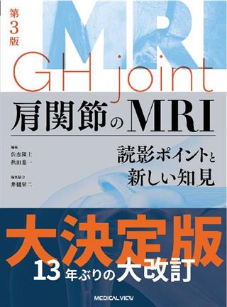
肩関節のMRI 読影ポイントと新しい知見 第3版

出版社: メジカルビュー社
著者:
発行日: 2024-04-01
分野: 臨床医学:一般  >  放射線/核医学
ISBN: 9784758321860
電子書籍版: 2024-04-01 (第3版第1刷)
書籍・雑誌
≪全国送料無料でお届け≫
取寄せ目安:8~14営業日

10,450 円(税込)

電子書籍
章別単位での購入はできません
ブラウザ、アプリ閲覧

10,450 円(税込)

商品紹介

定番書籍が13年を経て大改訂!
「凍結肩」の章を新たに設け50以上の症例を提示するほか,腫瘍類似性病変や神経疾患,新しいバイオメカニクスの知見も収載。MRI正常解剖のスライス数を増やし,各疾患の治療方針を解説,また特徴的な術後MRIを例示するなど,さらにわかりやすい紙面にアップグレード。読影に必要なMRI正常解剖も一新して,舌部の解剖も加りさらに充実!
これから学ぶ方にも,旧版をお持ちの方にも自信を持ってお薦めできる一冊。

目次

  • MRI正常解剖図譜
     斜位冠状断像,斜位矢状断像,軸位断像,舌部の正常解剖図譜

    1 基 礎
    肩関節の解剖とMRI
     肩関節の解剖を知る
     肩関節の層構造
     肩複合体(上肢帯)
     肩関節の骨
     肩の筋群
     肩関節上下方の安定化機構
     腱板疎部
     肩甲下滑液包
     肩峰下滑液包
     解剖写真でみる骨形態
     解剖写真でみる腱板の筋内腱,関節包の付着
    肩関節の機能解剖と病態
     肩関節の機能
     肩関節の病態
    肩関節のバイオメカニクス
     肩はどの関節よりも自由度が高いがそもそも不安定な関節
     肩は複数のテコが作用しておりバランスがとれていると腕を持ち上げることができる
    肩関節の外来診察
     外来診察
     新しい理学テスト
     疼痛肩
     スポーツ障害肩
     その他の肩関節疾患

    2 臨床
    疼痛肩-腱板断裂
     腱板とは
     腱板疎部とは
     腱板断裂とは
     棘上筋腱停止部,滑液包側剥離断裂の拡大様式
     棘上筋における外傷性筋腱間断裂とその拡大,および残存遠位端の萎縮消失
     棘上下筋腱全層断裂後に生じる“引き込み”と萎縮評価
     MRIで診断する腱板断裂
     MRIで診断する部分断裂
     MRIで診断する全層断裂
    疼痛肩-突き上げと擦れ(インピンジメント)
     中高年の疼痛肩
     突き上げと擦れ(インピンジメント)とは
    疼痛肩-石灰沈着性腱板炎
     石灰沈着性腱板炎とは
     MRIで診断する石灰沈着性腱板炎
    疼痛肩-上腕二頭筋長頭腱炎
     上腕二頭筋長頭腱炎とは
     MRIで診断する長頭腱炎
    疼痛肩-上腕二頭筋長頭腱断裂
     上腕二頭筋長頭腱断裂とは
     MRIで診断する長頭腱断裂
    疼痛肩-上腕二頭筋長頭腱亜脱臼,脱臼
     上腕二頭筋長頭腱亜脱臼,脱臼とは
     MRIで診断する長頭腱亜脱臼,脱臼
    疼痛肩-hidden lesion(隠された病変)―長頭腱滑車部での肩甲下筋腱舌部損傷―
     hidden lesionとは
     長頭腱亜脱臼を伴わないhidden lesionとは
     MRIで診断するhidden lesion
    疼痛肩-筋損傷
     肉ばなれとは
     筋挫傷とは
     MRIで診断する筋損傷
    疼痛肩-上腕骨頭大結節不全骨折,骨挫傷
     上腕骨頭大結節不全骨折,骨挫傷とは
     MRIで診断する上腕骨頭大結節不全骨折,骨挫傷
    疼痛肩-肩 関節包炎⊇拘縮肩(=凍結肩)
     拘縮肩とは
     腋窩嚢拘縮とは
     MRIで診断する腋窩嚢拘縮
     腱板疎部拘縮とは
     MRIで診断する腱板疎部拘縮
    疼痛肩-腱板疎部炎,腱板疎部損傷
     腱板疎部とは
     腱板疎部炎,腱板疎部損傷とは
     MRIで診断する腱板疎部炎,腱板疎部損傷
    疼痛肩-腋窩嚢(関節包)/関節唇の関節裂隙への手繰れ込み 
     手繰れ込みとは
    疼痛肩-凍結肩
     肩 関節包炎⊇凍結肩≒五十肩
     経過観察することができた肩 関節包炎
     合併症を認める肩 関節包炎
    スポーツ障害肩
     肩のスポーツ外傷とスポーツ障害
     投球障害肩
     後上方関節唇損傷(SLAP損傷)
     傍関節唇嚢胞(ガングリオン,paralabral cyst)
     関節内後方押し擦れ
     slant appearance,Bennett損傷
     小円筋の浮腫
     上腕骨近位骨端線離開
    不安定肩
     反復性肩関節脱臼
     動揺肩
    関節関連の腫瘍腫瘍類似疾患
     滑膜増殖性疾患
     関節近傍に生じる骨腫瘍
    その他の肩関節疾患
     変形性肩関節症(OA)
     化膿性肩関節炎
     リウマチ性肩関節症
     透析肩(hemodialysis-related shoulder)
     上腕骨頭壊死
     滑膜骨軟骨腫症(synovialosteochondromatosis)
     PVNS(pigmented villonodular synovitis;色素性絨毛結節性滑膜炎)
     肩の腫瘍,腫瘍類似性病変
     烏口下滑液包炎
     os acromiale
     BCG
     乳癌再発
     神経疾患

    3 MRIの基礎知識
    撮像方法
     検査の目的
     撮像方法を決める
     撮像方向を決める
     T1,T2,T2*,脂肪抑制のパルスシークエンスの選び方
     患者への対応
     撮像時の注意点
     ルーチン検査のプロトコル
     病変はどこにあるのか(位置決め撮像)
     MR-関節造影(MR-arthrography)

    読影の壺 疾患別にみる読影臨床のポイント
    疼痛肩
     腱板断裂⊇剥離
     腱板炎
     石灰沈着性腱板炎
     上腕二頭筋長頭腱炎
     上腕二頭筋長頭腱脱臼と肩甲下筋腱断裂
     肉ばなれ
     大結節骨折,骨挫傷
     肩 関節炎⊇関節周囲炎
     腱板疎部肩 関節包炎(凍結肩)
    スポーツ障害肩
     投球障害肩(上方〜後上方関節唇損傷,関節内後上方押し擦れ)
     投球障害肩(ガングリオン:関節唇損傷と傍関節唇嚢胞)
     投球障害肩(棘下筋腱,関節側,部分断裂腱挫傷)
     投球障害肩(棘下筋腱,関節側,剥離)
     腱板疎部炎
     腱板疎部損傷
    不安定肩
     狭義Bankart損傷,Hill-Sachs損傷
     骨性Bankart損傷,摩耗型骨性Bankart損傷
     HAGL
     動揺肩(loose shoulder)

    肩関節の整形外科用語

この書籍の参考文献

参考文献のリンクは、リンク先の都合等により正しく表示されない場合がありますので、あらかじめご了承下さい。

本参考文献は電子書籍掲載内容を元にしております。

1 基礎

P.33 掲載の参考文献
1) Clemente CD. Osteology : In Gray's Anatomy 30th American Edition. Lea & Febiger, Philadelphia, 1985, p.233-234.
2) Nozaki T, et al : The anatomic relationship between the morphology of the greater tubercle of the humerus and the insertion of the infraspinatus tendon. J Shoulder Elbow Surg, 24 : 555-560, 2015.
3) Arai R, et al : Subscapularis tendon tear : an anatomic and clinical investigation. Arthroscopy, 24 : 997-1004, 2008.
4) Mochizuki T, et al : Humeral Insertion of the supraspinatus and infraspinatus : New anatomical findings regarding the footprint of the rotator cuff. J Bone Joint Surg Am, 90 : 962-969, 2008.
5) Kato A, et al : An anatomical study of the transverse part of the infraspinatus muscle that is closely related with the supraspinatus muscle. Surg Radiol Anat, 34 : 257-265, 2012.
6) Hamada J, et al : Anatomic study and electromyographic analysis of the teres minor muscle. J Shoulder Elbow Surg, 26 : 870-877, 2017.
7) Nimura A, et al : The superior capsule of the shoulder joint complements the insertion of the rotator cuff. J Shoulder Elbow Surg, 21 : 867-872, 2012.
P.40 掲載の参考文献
1) Kazar B, Relovszky E : Prognosis of primary dislocation of the shoulder. Acta Orthop Scand, 40 : 216-224, 1969.
2) Poppen NK, Walker PS : Forces at the glenohumeral joint in abduction. Clin Orthop, 135 : 165-170, 1978.
3) 井樋栄二 : 肩学 : 臨床の「なぜ」とその追求. 医学書院, 東京, 2021.
4) Itoi E, et al : Scapular inclination and inferior stability of the shoulder. J Shoulder Elbow Surg, 1 : 131-139, 1992.
5) Itoi E, et al : Biomechanics of the shoulder. In : The Shoulder, 4th ed, Rockwood CA Jr, Matsen FA III, Wirth MA, Lippitt SB, eds. Saunders/Elsevier, Philadelphia, 2009, p213-265.
6) Warner JJ, McMahon PJ : The role of the long head of the biceps brachii in superior stability of theglenohumeral joint. J Bone Joint Surg Am, 77 : 366-372, 1995.
7) Kido T, et al : The depressor function of biceps on the head of the humerus in shoulders with tears of the rotator cuff. J Bone Joint Surg Br, 82 : 416-419, 2000.
8) Minagawa H, et al : Prevalence of symptomatic and asymptomatic rotator cuff tears in the general population : From mass-screening in one village. J Orthop, 10 : 8-12, 2013.
9) Yamamoto A, et al : Prevalence and risk factors of a rotator cuff tear in the general population. J Shoulder Elbow Surg, 19 : 116-120, 2010.
10) Itoi E, et al : The effect of a glenoid defect on anteroinferior stability of the shoulder after Bankart repair : a cadaveric study. J Bone Joint Surg Am, 82 : 35-46, 2000.
11) Yamamoto N, et al : Contact between the glenoid and the humeral head in abduction, external rotation, and horizontal extension : a new concept of glenoid track. J Shoulder Elbow Surg, 16 : 649-656, 2007.
12) Hovelius L, et al : Primary anterior dislocation of the shoulder in young patients : a ten-year prospective study. J Bone Joint Surg Am, 8 : 1677-1684, 1996.
13) Itoi E, et al : Position of immobilization after dislocation of the shoulder : a cadaveric study. J Bone Joint Surg Am, 81 : 385-390, 1999.
14) Itoi E, et al : Position of immobilization after dislocation of the glenohumeral joint : a study with use of magnetic resonance imaging. J Bone Joint Surg Am, 83 : 661-667, 2001.
15) Itoi E, et al : Immobilization in external rotation after shoulder dislocation reduces the risk of recurrence : a randomized controlled trial. J Bone Joint Surg Am, 89 : 2124-2131, 2007.
16) Shinagawa K, et al : Immobilization in External Rotation Reduces the Risk of Recurrence After Primary Anterior Shoulder Dislocation : A Metaanalysis. Orthop J Sports Med, 2020 Jun 15. [eCollection 2020 Jun]
17) Itoi E, et al : Long-Term Effect of Immobilization in External Rotation After First-Time Shoulder Dislocation : An Average 18-Year Follow-Up. J Shoulder Elbow Surg, 31 : 601-607, 2022.
18) Itoi E, et al : Arm abduction provides a better reduction of the Bankart lesion during immobilization in external rotation after an initial shoulder dislocation. Am J Sports Med, 43 : 1731-1736, 2015.
P.58 掲載の参考文献
1) Cnockaert JC, et al : Relative contribution of individual muscles to the isometric contraction of a muscular group. J Biomech, 8 : 191-197, 1975.
2) Matsubayashi Y, et al : Validity and reliability of the Japanese version of the pain DETECT questionnaire : a multicenter observational study. PLoS One, 30 : e68013, 2013.
3) Kim BK, et al : Flexion-extension-supination test compared to arthroscopic findings of biceps long head pathology : A physical examination that reflect anatomical evolution of human shoulder girdle. Medicine (Baltimore). 101 : e29755, 2022.
4) Rabin A, et al : Shoulder assessment by smartphone : a valid alternative for times of social distancing. Arch Orthop Trauma Surg, 142 : 979-985, 2022.
5) Wang G, et al ; Reliability and accuracy of telemedicine-based shoulder examinations. J Shoulder Elbow Surg, 31 : e369-e375, 2022.

2 臨床

P.121 掲載の参考文献
Malghem J, et al : Intraosseous migration of tendinous calcifications : cortical erosions, subcortical migration and extensive intramedullary diffusion, a SIMS series. Skeletal Radiol, 44 : 1403-1412, 2015.
Gwalani R, et al : MRI findings in intraosseous extension of calcific supraspinatus tendonitis. Radiol Case Rep, 15 : 975-977, 2020.
P.137 掲載の参考文献
1) 泉純一, ほか : 肩関節の "hidden lesion" のMRI所見. 画像診断, 22 : 662-667, 2002.
P.159 掲載の参考文献
1) 信原克哉 : 肩-その機能と臨床, 第2版. 医学書院, 東京, 1987.
2) 信原克哉 : 肩-その機能と臨床, 第3版. 医学書院, 東京, 2001.
P.275 掲載の参考文献
1) Lin J, et al : Pigmented villonodular synovitis and related lesions : the spectrum of imaging findings. AJR Am J Roentgenol, 172 : 191-197, 1999.
2) Ryu KN, et al : MR imaging of lipoma arborescens of the knee joint AJR Am J Roentgenol, 167 : 1229-1232, 1996.
3) Kramer J, et al : MR appearance of idiopathic synovial osteo-chondromatosis. J Comput Assist Tomogr, 17 : 772-776, 1993.
4) Greenspan A, et al : Synovial hemangioma : imaging features in eight histologically proven cases, review of the literature, and differential diagnosis. Skeletal Radiol, 24 : 583-590, 1995.
5) Davies M, et al : The diagnostic accuracy of MR imaging in osteoid osteoma. Skeletal Radiol, 10 : 559-569, 2002.
P.295 掲載の参考文献
1) 高松勇, ほか : 最近のBCG接種の効果をめぐって. 結核, 70 : 561-564, 1995.
2) 森亨 : 新しいBCG接種制度とBCG接種の副反応. 小児科, 48 : 2011-2017, 2007.
3) 第41回乳癌研究会 : 乳癌再発後生存期間に関するアンケート基本集計. 乳癌研究会, 名古屋, 1985, p28.
4) 唐司則之 : 乳癌鎖骨上窩リンパ節再発例に対する治療法の検討. 乳癌の臨床, 7 : 541-544, 1992.
5) Nascimento AB, et al : Diffuse desmoplastic breast carcinoma metastases to the liver simulating cirrhosis at MR imaging : report of two cases. Radiology, 221 : 117-121, 2001.

最近チェックした商品履歴

Loading...